 Movie
Movie Controller

[English] 日本語
 Yorodumi
- PDB-1te5: The 2.0 Angstrom crystal structure of predicted glutamine amidotr... -
+ Open data

- Basic information
| Entry | Database: PDB / ID: 1te5 | ||||||
|---|---|---|---|---|---|---|---|
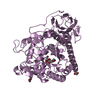
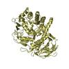
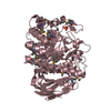
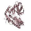
| Title | The 2.0 Angstrom crystal structure of predicted glutamine amidotransferase from Pseudomonas aeruginosa PA01 | ||||||
 Components Components | conserved hypothetical protein | ||||||
 Keywords Keywords | UNKNOWN FUNCTION / glutamine amidotransferase / amidotransferase / Structural Genomics / PSI / Protein Structure Initiative / New York SGX Research Center for Structural Genomics / NYSGXRC | ||||||
| Function / homology |  Function and homology information Function and homology informationGamma-glutamyl-hercynylcysteine sulfoxide hydrolase EgtC-like / Glutamine amidotransferases class-II / Glutamine amidotransferase type 2 domain profile. / Glutamine amidotransferase type 2 domain / Aminohydrolase, N-terminal nucleophile (Ntn) domain / Glutamine Phosphoribosylpyrophosphate, subunit 1, domain 1 / Nucleophile aminohydrolases, N-terminal / 4-Layer Sandwich / Alpha Beta Similarity search - Domain/homology | ||||||
| Biological species |  Pseudomonas aeruginosa PAO1 (bacteria) Pseudomonas aeruginosa PAO1 (bacteria) | ||||||
| Method |  X-RAY DIFFRACTION / MIR / Resolution: 2 Å X-RAY DIFFRACTION / MIR / Resolution: 2 Å | ||||||
 Authors Authors | Patskovsky, Y. / Almo, S.C. / Burley, S.K. / New York SGX Research Center for Structural Genomics (NYSGXRC) | ||||||
 Citation Citation | Journal: To be Published Title: Crystal structure of amidophosphoribosyltransferase from Pseudomonas aeruginosa Authors: Patskovsky, Y. / Almo, S.C. | ||||||
| History |
| ||||||
| Remark 300 | BIOMOLECULE: THIS ENTRY CONTAINS THE CRYSTALLOGRAPHIC ASYMMETRIC UNIT WHICH CONSISTS OF 2 CHAIN(S). ...BIOMOLECULE: THIS ENTRY CONTAINS THE CRYSTALLOGRAPHIC ASYMMETRIC UNIT WHICH CONSISTS OF 2 CHAIN(S). THE BIOLOGICAL UNIT IS UNKNOWN. |
- Structure visualization
| Structure viewer | Molecule: MolmilJmol/JSmol |
|---|
- Downloads & links
-Download
| PDBx/mmCIF format | 1te5.cif.gz | 112.8 KB | Display | PDBx/mmCIF format |
|---|---|---|---|---|
| PDB format | pdb1te5.ent.gz | 87.6 KB | Display | PDB format |
| PDBx/mmJSON format | 1te5.json.gz | Tree view | PDBx/mmJSON format | |
| Others |  Other downloads Other downloads |
-Validation report
| Arichive directory | https://data.pdbj.org/pub/pdb/validation_reports/te/1te5ftp://data.pdbj.org/pub/pdb/validation_reports/te/1te5 | HTTPS FTP |
|---|
-Related structure data
| Similar structure data | |
|---|---|
| Other databases |


-Links
 PDBj
PDBj


- Assembly
| Deposited unit | 
| ||||||||
|---|---|---|---|---|---|---|---|---|---|
| 1 |
| ||||||||
| Unit cell |
|
-Components
| #1: Protein | Mass: 28575.387 Da / Num. of mol.: 2 Source method: isolated from a genetically manipulated source Source: (gene. exp.) Pseudomonas aeruginosa PAO1 (bacteria) / Species: Pseudomonas aeruginosa / Strain: PAO1 / Gene: PA1307 / Plasmid: pET / Species (production host): Escherichia coli / Production host: #2: Water | ChemComp-HOH / |  Mass: 18.015 Da / Num. of mol.: 191 / Source method: isolated from a natural source / Formula: H2O Mass: 18.015 Da / Num. of mol.: 191 / Source method: isolated from a natural source / Formula: H2O |
|---|
-Experimental details
-Experiment
| Experiment | Method: X-RAY DIFFRACTION / Number of used crystals: 2 |
|---|
- Sample preparation
| Crystal |
| |||||||||
|---|---|---|---|---|---|---|---|---|---|---|
| Crystal grow | Temperature: 280 K / pH: 7.5 Details: 0.1M HEPES, 28% PEG 3350, pH 7.5, VAPOR DIFFUSION, SITTING DROP, temperature 280K, pH 7.50 |
-Data collection
| Diffraction |
| |||||||||||||||
|---|---|---|---|---|---|---|---|---|---|---|---|---|---|---|---|---|
| Diffraction source | Source: ROTATING ANODE / Type: RIGAKU RU200 / Wavelength: 1.5418 | |||||||||||||||
| Detector |
| |||||||||||||||
| Radiation | Monochromator: GRAPHITE / Protocol: SINGLE WAVELENGTH / Monochromatic (M) / Laue (L): M / Scattering type: x-ray | |||||||||||||||
| Radiation wavelength | Wavelength: 1.5418 Å / Relative weight: 1 | |||||||||||||||
| Reflection | Resolution: 2→20 Å / Num. obs: 32917 / % possible obs: 87.8 % / Observed criterion σ(I): 0 / Redundancy: 29.4 % / Biso Wilson estimate: 13.6 Å2 / Rmerge(I) obs: 0.079 / Rsym value: 0.079 / Net I/σ(I): 9.7 | |||||||||||||||
| Reflection shell | Resolution: 2→2.07 Å / Redundancy: 4.3 % / Rmerge(I) obs: 0.26 / Mean I/σ(I) obs: 2.2 / Rsym value: 0.32 / % possible all: 43.6 |
- Processing
| Software |
| ||||||||||||||||||||||||||||||||||||||||||||||||||||||||||||||||||||||||||||||||
|---|---|---|---|---|---|---|---|---|---|---|---|---|---|---|---|---|---|---|---|---|---|---|---|---|---|---|---|---|---|---|---|---|---|---|---|---|---|---|---|---|---|---|---|---|---|---|---|---|---|---|---|---|---|---|---|---|---|---|---|---|---|---|---|---|---|---|---|---|---|---|---|---|---|---|---|---|---|---|---|---|---|
| Refinement | Method to determine structure: MIR / Resolution: 2→20 Å / Rfactor Rfree error: 0.009 / Data cutoff high absF: 300000 / Data cutoff low absF: 0.01 / Isotropic thermal model: ISOTROPIC / Cross valid method: THROUGHOUT / σ(F): 1 / Stereochemistry target values: ENGH & HUBER / Details: USED WEIGHTED FULL MATRIX LEAST SQUARES PROCEDURE
| ||||||||||||||||||||||||||||||||||||||||||||||||||||||||||||||||||||||||||||||||
| Solvent computation | Solvent model: FLAT MODEL / Bsol: 28.9525 Å2 / ksol: 0.319038 e/Å3 | ||||||||||||||||||||||||||||||||||||||||||||||||||||||||||||||||||||||||||||||||
| Displacement parameters | Biso mean: 39 Å2
| ||||||||||||||||||||||||||||||||||||||||||||||||||||||||||||||||||||||||||||||||
| Refine analyze |
| ||||||||||||||||||||||||||||||||||||||||||||||||||||||||||||||||||||||||||||||||
| Refinement step | Cycle: LAST / Resolution: 2→20 Å
| ||||||||||||||||||||||||||||||||||||||||||||||||||||||||||||||||||||||||||||||||
| Refine LS restraints |
| ||||||||||||||||||||||||||||||||||||||||||||||||||||||||||||||||||||||||||||||||
| LS refinement shell | Resolution: 2→2.13 Å / Rfactor Rfree error: 0.038 / Total num. of bins used: 6
| ||||||||||||||||||||||||||||||||||||||||||||||||||||||||||||||||||||||||||||||||
| Xplor file |
|
