 Movie
Movie Controller

[English] 日本語
 Yorodumi
- PDB-1t6o: Nucleocapsid-binding domain of the measles virus P protein (amino... -
+ Open data

- Basic information
| Entry | Database: PDB / ID: 1t6o | ||||||
|---|---|---|---|---|---|---|---|
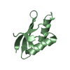
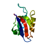
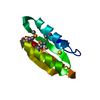
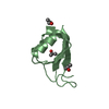
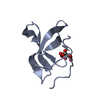
| Title | Nucleocapsid-binding domain of the measles virus P protein (amino acids 457-507) in complex with amino acids 486-505 of the measles virus N protein | ||||||
 Components Components |
| ||||||
 Keywords Keywords |  VIRAL PROTEIN / four helix bundle VIRAL PROTEIN / four helix bundle | ||||||
| Function / homology |  Function and homology information Function and homology informationhelical viral capsid / viral genome replication / viral nucleocapsid / host cell cytoplasm / molecular adaptor activity / ribonucleoprotein complex / RNA-dependent RNA polymerase activity / DNA-templated transcription / structural molecule activity / RNA bindingSimilarity search - Function | ||||||
| Biological species |   Measles virus Measles virus | ||||||
| Method | X-RAY DIFFRACTION / MOLECULAR REPLACEMENT / Resolution: 2 Å | ||||||
 Authors Authors | Kingston, R.L. / Hamel, D.J. / Gay, L.S. / Dahlquist, F.W. / Matthews, B.W. | ||||||
 Citation Citation | Journal: Proc.Natl.Acad.Sci.USA / Year: 2004 Title: Structural basis for the attachment of a paramyxoviral polymerase to its template. Authors: Kingston, R.L. / Hamel, D.J. / Gay, L.S. / Dahlquist, F.W. / Matthews, B.W. #1: Journal: To be PublishedTitle: Characterization of nucleocapsid binding by the measles and the mumps virus phosphoprotein Authors: Kingston, R.L. / Baase, W.A. / Gay, L.S. | ||||||
| History |
| ||||||
| Remark 999 | SEQUENCE Structure of a chimeric molecule encompassing amino acids 457-507 of measles P and 486-505 ...SEQUENCE Structure of a chimeric molecule encompassing amino acids 457-507 of measles P and 486-505 of measles N. They are connected by a flexible 8 amino acid linker (GS)4, which is largely disordered in the crystal structure. The asymmetric unit of the crystal contains the P moiety from one molecule (chain A) bound to the N moiety of a different molecule (Chain B). Link record between chain A and L is not provided since the part of the chain L which is linked to chain A is missing from the coordinates due to lack of electron density. |
- Structure visualization
| Structure viewer | Molecule: MolmilJmol/JSmol |
|---|
- Downloads & links
-Download
| PDBx/mmCIF format | 1t6o.cif.gz | 26.3 KB | Display | PDBx/mmCIF format |
|---|---|---|---|---|
| PDB format | pdb1t6o.ent.gz | 16.2 KB | Display | PDB format |
| PDBx/mmJSON format | 1t6o.json.gz | Tree view | PDBx/mmJSON format | |
| Others |  Other downloads Other downloads |
-Validation report
| Arichive directory | https://data.pdbj.org/pub/pdb/validation_reports/t6/1t6oftp://data.pdbj.org/pub/pdb/validation_reports/t6/1t6o | HTTPS FTP |
|---|
-Related structure data
| Related structure data | 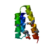 1oksS S: Starting model for refinement |
|---|---|
| Similar structure data |


-Links
 PDBj
PDBj

- Assembly
| Deposited unit | 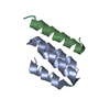
| ||||||||
|---|---|---|---|---|---|---|---|---|---|
| 1 |
| ||||||||
| Unit cell |
|
-Components
| #1: Protein | Mass: 5891.055 Da / Num. of mol.: 1 / Fragment: residues 457-507 / Mutation: P458G Source method: isolated from a genetically manipulated source Details: Chimeric molecule encompassing amino acids 457-507 of measles P (chain A) and 486-505 of measles N (chain B). They are connected by a flexible 8 amino acid linker (GS)4 (chain L) Source: (gene. exp.) Measles virus / Genus: Morbillivirus / Strain: Moraten vaccine strain / Gene: P/V / Plasmid: pET41a(+) / Production host:  Escherichia coli (E. coli) / Strain (production host): BL21-Star(DE3) / References: GenBank: 9181875, UniProt: Q77M42*PLUS Escherichia coli (E. coli) / Strain (production host): BL21-Star(DE3) / References: GenBank: 9181875, UniProt: Q77M42*PLUS |
|---|---|
| #2: Protein/peptide | Mass: 594.534 Da / Num. of mol.: 1 / Source method: obtained synthetically / Details: synthetic linker |
| #3: Protein/peptide | Mass: 2162.453 Da / Num. of mol.: 1 / Fragment: residues 486-505 Source method: isolated from a genetically manipulated source Source: (gene. exp.) Measles virus / Genus: Morbillivirus / Strain: Moraten vaccine strain / Gene: N / Plasmid: pET41a(+) / Production host: Escherichia coli (E. coli) / Strain (production host): BL21-Star(DE3) / References: GenBank: 9181874, UniProt: Q77M43*PLUS |
| #4: Water | ChemComp-HOH / Water Mass: 18.015 Da / Num. of mol.: 28 / Source method: isolated from a natural source / Formula: H2O Mass: 18.015 Da / Num. of mol.: 28 / Source method: isolated from a natural source / Formula: H2O |
-Experimental details
-Experiment
| Experiment | Method: X-RAY DIFFRACTION / Number of used crystals: 1 |
|---|
- Sample preparation
| Crystal | Density Matthews: 2.11 Å3/Da / Density % sol: 41.64 % |
|---|---|
| Crystal grow | Temperature: 296 K / Method: vapor diffusion / pH: 9.1 Details: 0.2M AMPSO/KOH buffer, 0.5 - 1.0 M Ammonium sulfate, pH 9.1, VAPOR DIFFUSION, temperature 296K |
-Data collection
| Diffraction | Mean temperature: 298 K |
|---|---|
| Diffraction source | Source: ROTATING ANODE / Type: RIGAKU / Wavelength: 1.5418 Å |
| Detector | Type: RIGAKU RAXIS IV / Detector: IMAGE PLATE / Date: Mar 4, 2004 |
| Radiation | Monochromator: Graphite / Protocol: SINGLE WAVELENGTH / Monochromatic (M) / Laue (L): M / Scattering type: x-ray |
| Radiation wavelength | Wavelength: 1.5418 Å / Relative weight: 1 |
| Reflection | Resolution: 2→25 Å / Num. all: 5306 / Num. obs: 5306 / % possible obs: 97.5 % / Observed criterion σ(F): 0 / Observed criterion σ(I): 0 |
| Reflection shell | Resolution: 2→2.03 Å / % possible all: 96.4 |
- Processing
| Software |
| |||||||||||||||||||||||||
|---|---|---|---|---|---|---|---|---|---|---|---|---|---|---|---|---|---|---|---|---|---|---|---|---|---|---|
| Refinement | Method to determine structure: MOLECULAR REPLACEMENT Starting model: 1OKS.pdb Resolution: 2→25 Å / Isotropic thermal model: Individual Isotropic B / Cross valid method: test set omitted from all refinement / σ(F): 0 / σ(I): 0 / Stereochemistry target values: Engh & Huber
| |||||||||||||||||||||||||
| Solvent computation | Solvent model: Moews & Kretsinger / Bsol: 300 Å2 / ksol: 0.85 e/Å3 | |||||||||||||||||||||||||
| Displacement parameters |
| |||||||||||||||||||||||||
| Refinement step | Cycle: LAST / Resolution: 2→25 Å
| |||||||||||||||||||||||||
| Refine LS restraints |
| |||||||||||||||||||||||||
| Xplor file |
|
