 Movie
Movie Controller

[English] 日本語
 Yorodumi
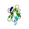
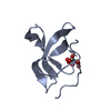
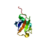
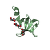
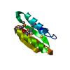
- PDB-1osd: crystal structure of Oxidized MerP from Ralstonia metallidurans CH34 -
+ Open data

- Basic information
| Entry | Database: PDB / ID: 1osd | ||||||
|---|---|---|---|---|---|---|---|
| Title | crystal structure of Oxidized MerP from Ralstonia metallidurans CH34 | ||||||
 Components Components | hypothetical protein MerP | ||||||
 Keywords Keywords | METAL BINDING PROTEIN / Mercury resistance / Perisplasm / structural genomics | ||||||
| Function / homology | Alpha-Beta Plaits - #100 / Alpha-Beta Plaits / 2-Layer Sandwich / Alpha Beta / :  Function and homology information Function and homology information | ||||||
| Biological species |  Cupriavidus metallidurans (bacteria) Cupriavidus metallidurans (bacteria) | ||||||
| Method |  X-RAY DIFFRACTION / MOLECULAR REPLACEMENT / Resolution: 2 Å X-RAY DIFFRACTION / MOLECULAR REPLACEMENT / Resolution: 2 Å | ||||||
 Authors Authors | Serre, L. / Rossy, E. / Pebay-Peyroula, E. / Cohen-Addad, C. / Coves, J. | ||||||
 Citation Citation | Journal: J.MOL.BIOL. / Year: 2004 Title: Crystal Structure of the Oxidized Form of the Periplasmic Mercury-binding Protein MerP from Ralstonia metallidurans CH34 Authors: Serre, L. / Rossy, E. / Pebay-Peyroula, E. / Cohen-Addad, C. / Coves, J. | ||||||
| History |
|
- Structure visualization
| Structure viewer | Molecule: MolmilJmol/JSmol |
|---|
- Downloads & links
-Download
| PDBx/mmCIF format | 1osd.cif.gz | 41.1 KB | Display | PDBx/mmCIF format |
|---|---|---|---|---|
| PDB format | pdb1osd.ent.gz | 29.3 KB | Display | PDB format |
| PDBx/mmJSON format | 1osd.json.gz | Tree view | PDBx/mmJSON format | |
| Others |  Other downloads Other downloads |
-Validation report
| Arichive directory | https://data.pdbj.org/pub/pdb/validation_reports/os/1osdftp://data.pdbj.org/pub/pdb/validation_reports/os/1osd | HTTPS FTP |
|---|
-Related structure data
| Related structure data | 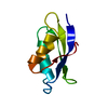 1afiS S: Starting model for refinement |
|---|---|
| Similar structure data |


-Links
 PDBj
PDBj- Assembly
| Deposited unit | 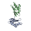
| ||||||||||||||||||
|---|---|---|---|---|---|---|---|---|---|---|---|---|---|---|---|---|---|---|---|
| 1 | 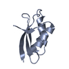
| ||||||||||||||||||
| 2 | 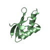
| ||||||||||||||||||
| Unit cell |
| ||||||||||||||||||
| Noncrystallographic symmetry (NCS) | NCS domain:
NCS domain segments: Component-ID: 1 / Ens-ID: 1 / Beg auth comp-ID: ALA / Beg label comp-ID: ALA / End auth comp-ID: VAL / End label comp-ID: VAL / Refine code: 5 / Auth seq-ID: 1 - 70 / Label seq-ID: 1 - 70
| ||||||||||||||||||
| Details | The biological assembly is a monomer |
-Components
| #1: Protein | Mass: 7530.622 Da / Num. of mol.: 2 / Fragment: residues 1-72 Source method: isolated from a genetically manipulated source Source: (gene. exp.) Cupriavidus metallidurans (bacteria) / Gene: MerP / Plasmid: pET-3a / Species (production host): Escherichia coli / Production host: #2: Water | ChemComp-HOH / |  Mass: 18.015 Da / Num. of mol.: 125 / Source method: isolated from a natural source / Formula: H2O Mass: 18.015 Da / Num. of mol.: 125 / Source method: isolated from a natural source / Formula: H2OHas protein modification | Y | |
|---|
-Experimental details
-Experiment
| Experiment | Method: X-RAY DIFFRACTION / Number of used crystals: 1 |
|---|
- Sample preparation
| Crystal | Density Matthews: 2.03 Å3/Da / Density % sol: 39.02 % |
|---|---|
| Crystal grow | Temperature: 293 K / Method: vapor diffusion, hanging drop / pH: 8.5 Details: PEGMME5000, Sodium acetate, Tris-HCl, pH 8.5, VAPOR DIFFUSION, HANGING DROP, temperature 293K |
-Data collection
| Diffraction | Mean temperature: 100 K |
|---|---|
| Diffraction source | Source: ROTATING ANODE / Type: ENRAF-NONIUS FR591 / Wavelength: 1.5418 Å |
| Detector | Type: MARRESEARCH / Detector: IMAGE PLATE / Details: osmic multi-layer mirror |
| Radiation | Protocol: SINGLE WAVELENGTH / Monochromatic (M) / Laue (L): M / Scattering type: x-ray |
| Radiation wavelength | Wavelength: 1.5418 Å / Relative weight: 1 |
| Reflection | Resolution: 2→40 Å / Num. obs: 9437 / % possible obs: 97 % / Observed criterion σ(F): 1 / Observed criterion σ(I): 0 / Redundancy: 4.2 % / Biso Wilson estimate: 35 Å2 / Rmerge(I) obs: 0.054 / Rsym value: 0.048 / Net I/σ(I): 10.7 |
| Reflection shell | Resolution: 2→2.05 Å / Redundancy: 3.7 % / Rmerge(I) obs: 0.335 / Mean I/σ(I) obs: 2.1 / Num. unique all: 655 / Rsym value: 0.282 / % possible all: 97 |
- Processing
| Software |
| ||||||||||||||||||||||||||||||||||||||||||||||||||||||||||||||||||||||||||||||||||||||||||||||||||||
|---|---|---|---|---|---|---|---|---|---|---|---|---|---|---|---|---|---|---|---|---|---|---|---|---|---|---|---|---|---|---|---|---|---|---|---|---|---|---|---|---|---|---|---|---|---|---|---|---|---|---|---|---|---|---|---|---|---|---|---|---|---|---|---|---|---|---|---|---|---|---|---|---|---|---|---|---|---|---|---|---|---|---|---|---|---|---|---|---|---|---|---|---|---|---|---|---|---|---|---|---|---|
| Refinement | Method to determine structure: MOLECULAR REPLACEMENT Starting model: PDB entry 1AFI Resolution: 2→39.53 Å / Cor.coef. Fo:Fc: 0.954 / Cor.coef. Fo:Fc free: 0.919 / SU B: 5.418 / SU ML: 0.15 / Cross valid method: THROUGHOUT / σ(F): 0 / ESU R: 0.233 / ESU R Free: 0.212 / Stereochemistry target values: MAXIMUM LIKELIHOOD / Details: HYDROGENS HAVE BEEN ADDED IN THE RIDING POSITIONS
| ||||||||||||||||||||||||||||||||||||||||||||||||||||||||||||||||||||||||||||||||||||||||||||||||||||
| Solvent computation | Ion probe radii: 0.8 Å / Shrinkage radii: 0.8 Å / VDW probe radii: 1.4 Å / Solvent model: BABINET MODEL WITH MASK | ||||||||||||||||||||||||||||||||||||||||||||||||||||||||||||||||||||||||||||||||||||||||||||||||||||
| Displacement parameters | Biso mean: 33.569 Å2
| ||||||||||||||||||||||||||||||||||||||||||||||||||||||||||||||||||||||||||||||||||||||||||||||||||||
| Refinement step | Cycle: LAST / Resolution: 2→39.53 Å
| ||||||||||||||||||||||||||||||||||||||||||||||||||||||||||||||||||||||||||||||||||||||||||||||||||||
| Refine LS restraints |
| ||||||||||||||||||||||||||||||||||||||||||||||||||||||||||||||||||||||||||||||||||||||||||||||||||||
| Refine LS restraints NCS | Dom-ID: 1 / Auth asym-ID: A / Ens-ID: 1 / Refine-ID: X-RAY DIFFRACTION
| ||||||||||||||||||||||||||||||||||||||||||||||||||||||||||||||||||||||||||||||||||||||||||||||||||||
| LS refinement shell | Resolution: 2→2.052 Å / Total num. of bins used: 20 /
|
