 Movie
Movie Controller


+ Open data

- Basic information
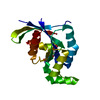
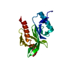
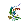
| Entry | Database: PDB / ID: 1shx | |||||||||
|---|---|---|---|---|---|---|---|---|---|---|
| Title | Ephrin A5 ligand structure | |||||||||
 Components Components | Ephrin-A5 Ephrin A5 Ephrin A5 | |||||||||
 Keywords Keywords | HORMONE/GROWTH FACTOR / ephrin signaling / HORMONE-GROWTH FACTOR COMPLEX | |||||||||
| Function / homology |  Function and homology information Function and homology informationneurotrophin TRKB receptor binding / EPH-Ephrin signaling / EPHA-mediated growth cone collapse / EPH-ephrin mediated repulsion of cells / negative regulation of substrate adhesion-dependent cell spreading / synaptic membrane adhesion / regulation of cell-cell adhesion / collateral sprouting / cellular response to follicle-stimulating hormone stimulus / positive regulation of collateral sprouting ...neurotrophin TRKB receptor binding / EPH-Ephrin signaling / EPHA-mediated growth cone collapse / EPH-ephrin mediated repulsion of cells / negative regulation of substrate adhesion-dependent cell spreading / synaptic membrane adhesion / regulation of cell-cell adhesion / collateral sprouting / cellular response to follicle-stimulating hormone stimulus / positive regulation of collateral sprouting / regulation of insulin secretion involved in cellular response to glucose stimulus / chemorepellent activity / regulation of cell morphogenesis / retinal ganglion cell axon guidance / positive regulation of synapse assembly / regulation of focal adhesion assembly / regulation of GTPase activity / basement membrane / GABA-ergic synapse / ephrin receptor signaling pathway / regulation of microtubule cytoskeleton organization / cellular response to forskolin / ephrin receptor binding / caveola / cell periphery / axon guidance / regulation of actin cytoskeleton organization / adherens junction / positive regulation of peptidyl-tyrosine phosphorylation / external side of plasma membrane / plasma membraneSimilarity search - Function | |||||||||
| Biological species |  Mus musculus (house mouse) Mus musculus (house mouse) | |||||||||
| Method | X-RAY DIFFRACTION / SYNCHROTRON / MOLECULAR REPLACEMENT / Resolution: 2.1 Å | |||||||||
 Authors Authors | Himanen, J.P. / Barton, W.A. / Nikolov, D.B. / Jeffrey, P.D. | |||||||||
 Citation Citation | Journal: J.Biol.Chem. / Year: 2005 Title: Three distinct molecular surfaces in ephrin-A5 are essential for a functional interaction with EphA3. Authors: Day, B. / To, C. / Himanen, J.P. / Smith, F.M. / Nikolov, D.B. / Boyd, A.W. / Lackmann, M. #1: Journal: Nat.Neurosci. / Year: 2004 Title: Repelling class discrimination: ephrin-A5 binds to and activates EphB2 receptor signaling. Authors: Himanen, J.P. / Chumley, M.J. / Lackmann, M. / Li, C. / Barton, W.A. / Jeffrey, P.D. / Vearing, C. / Geleick, D. / Feldheim, D.A. / Boyd, A.W. / Henkemeyer, M. / Nikolov, D.B. | |||||||||
| History |
|
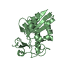
- Structure visualization
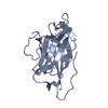
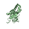
| Structure viewer | Molecule: MolmilJmol/JSmol |
|---|
- Downloads & links
-Download
| PDBx/mmCIF format | 1shx.cif.gz | 73.9 KB | Display | PDBx/mmCIF format |
|---|---|---|---|---|
| PDB format | pdb1shx.ent.gz | 55.6 KB | Display | PDB format |
| PDBx/mmJSON format | 1shx.json.gz | Tree view | PDBx/mmJSON format | |
| Others |  Other downloads Other downloads |
-Validation report
| Arichive directory | https://data.pdbj.org/pub/pdb/validation_reports/sh/1shxftp://data.pdbj.org/pub/pdb/validation_reports/sh/1shx | HTTPS FTP |
|---|
-Related structure data
| Related structure data | |
|---|---|
| Similar structure data |


-Links
 PDBj
PDBj
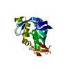
- Assembly
| Deposited unit | 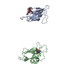
| ||||||||
|---|---|---|---|---|---|---|---|---|---|
| 1 | 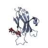
| ||||||||
| 2 | 
| ||||||||
| Unit cell |
|
-Components
| #1: Protein | Ephrin A5 / EPH-related receptor tyrosine kinase ligand 7 / LERK-7 / AL-1 Mass: 16320.332 Da / Num. of mol.: 2 Source method: isolated from a genetically manipulated source Source: (gene. exp.) Mus musculus (house mouse) / Gene: EFNA5, EPLG7, LERK7, EPL7 / Cell line (production host): HEK293 / Organ (production host): kidney / Production host:  Homo sapiens (human) / References: UniProt: O08543 Homo sapiens (human) / References: UniProt: O08543#2: Polysaccharide | 2-acetamido-2-deoxy-beta-D-glucopyranose-(1-4)-2-acetamido-2-deoxy-beta-D-glucopyranose-(1-4)-2- ...2-acetamido-2-deoxy-beta-D-glucopyranose-(1-4)-2-acetamido-2-deoxy-beta-D-glucopyranose-(1-4)-2-acetamido-2-deoxy-beta-D-glucopyranose / triacetyl-beta-chitotriose |   , Oligosaccharide / Class: Inhibitor / Mass: 627.594 Da / Num. of mol.: 1 , Oligosaccharide / Class: Inhibitor / Mass: 627.594 Da / Num. of mol.: 1Source method: isolated from a genetically manipulated source Details: oligosaccharide / References: triacetyl-beta-chitotriose #3: Polysaccharide | 2-acetamido-2-deoxy-beta-D-glucopyranose-(1-4)-2-acetamido-2-deoxy-beta-D-glucopyranose | / Mass: 424.401 Da / Num. of mol.: 1Source method: isolated from a genetically manipulated source #4: Water | ChemComp-HOH / | Water Mass: 18.015 Da / Num. of mol.: 105 / Source method: isolated from a natural source / Formula: H2O Mass: 18.015 Da / Num. of mol.: 105 / Source method: isolated from a natural source / Formula: H2O |
|---|
-Experimental details
-Experiment
| Experiment | Method: X-RAY DIFFRACTION / Number of used crystals: 1 |
|---|
- Sample preparation
| Crystal | Density Matthews: 2.26 Å3/Da / Density % sol: 45.58 % |
|---|---|
| Crystal grow | Temperature: 278 K / Method: vapor diffusion, hanging drop / pH: 4.6 Details: PEG 3350, pH 4.6, VAPOR DIFFUSION, HANGING DROP, temperature 278K |
-Data collection
| Diffraction | Mean temperature: 100 K |
|---|---|
| Diffraction source | Source: SYNCHROTRON / Site: NSLS  / Beamline: X9A / Wavelength: 0.979 Å / Beamline: X9A / Wavelength: 0.979 Å |
| Detector | Type: MARRESEARCH / Detector: CCD / Date: Dec 1, 2003 |
| Radiation | Protocol: SINGLE WAVELENGTH / Monochromatic (M) / Laue (L): M / Scattering type: x-ray |
| Radiation wavelength | Wavelength: 0.979 Å / Relative weight: 1 |
| Reflection | Resolution: 2→20 Å / Num. all: 17042 / Num. obs: 16487 / % possible obs: 91.3 % / Observed criterion σ(F): 0 / Observed criterion σ(I): 0 / Net I/σ(I): 16.5 |
| Reflection shell | Resolution: 2→2.09 Å / Rmerge(I) obs: 0.056 / Num. unique all: 17958 / % possible all: 54.8 |
- Processing
| Software |
| ||||||||||||||||||||
|---|---|---|---|---|---|---|---|---|---|---|---|---|---|---|---|---|---|---|---|---|---|
| Refinement | Method to determine structure: MOLECULAR REPLACEMENT Starting model: EprinB2 Resolution: 2.1→8 Å / σ(F): 0 / Stereochemistry target values: Engh & Huber
| ||||||||||||||||||||
| Refinement step | Cycle: LAST / Resolution: 2.1→8 Å
| ||||||||||||||||||||
| Refine LS restraints |
|
