 Movie
Movie Controller

[English] 日本語
 Yorodumi
- PDB-1rwz: Crystal Structure of Proliferating Cell Nuclear Antigen (PCNA) fr... -
+ Open data

- Basic information
| Entry | Database: PDB / ID: 1rwz | ||||||
|---|---|---|---|---|---|---|---|
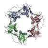
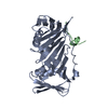
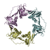
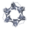
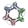
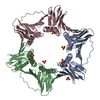
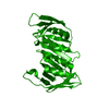
| Title | Crystal Structure of Proliferating Cell Nuclear Antigen (PCNA) from A. fulgidus | ||||||
 Components Components | DNA polymerase sliding clamp | ||||||
 Keywords Keywords | REPLICATION / sliding clamp / torus / processivity factor | ||||||
| Function / homology |  Function and homology information Function and homology informationDNA polymerase processivity factor activity / leading strand elongation / regulation of DNA replication / mismatch repair / translesion synthesis / DNA binding Similarity search - Function | ||||||
| Biological species |   Archaeoglobus fulgidus (archaea) Archaeoglobus fulgidus (archaea) | ||||||
| Method |  X-RAY DIFFRACTION / SYNCHROTRON / MOLECULAR REPLACEMENT / Resolution: 1.8 Å X-RAY DIFFRACTION / SYNCHROTRON / MOLECULAR REPLACEMENT / Resolution: 1.8 Å | ||||||
 Authors Authors | Chapados, B.R. / Hosfield, D.J. / Han, S. / Qiu, J. / Yelent, B. / Shen, B. / Tainer, J.A. | ||||||
 Citation Citation | Journal: Cell(Cambridge,Mass.) / Year: 2004 Title: Structural Basis for FEN-1 Substrate Specificity and PCNA-Mediated Activation in DNA Replication and Repair Authors: Chapados, B.R. / Hosfield, D.J. / Han, S. / Qiu, J. / Yelent, B. / Shen, B. / Tainer, J.A. | ||||||
| History |
|
- Structure visualization
| Structure viewer | Molecule: MolmilJmol/JSmol |
|---|
- Downloads & links
-Download
| PDBx/mmCIF format | 1rwz.cif.gz | 66.3 KB | Display | PDBx/mmCIF format |
|---|---|---|---|---|
| PDB format | pdb1rwz.ent.gz | 51.1 KB | Display | PDB format |
| PDBx/mmJSON format | 1rwz.json.gz | Tree view | PDBx/mmJSON format | |
| Others |  Other downloads Other downloads |
-Validation report
| Summary document | 1rwz_validation.pdf.gz | 424.6 KB | Display | wwPDB validaton report |
|---|---|---|---|---|
| Full document | 1rwz_full_validation.pdf.gz | 429.1 KB | Display | |
| Data in XML | 1rwz_validation.xml.gz | 14.4 KB | Display | |
| Data in CIF | 1rwz_validation.cif.gz | 21.4 KB | Display | |
| Arichive directory | https://data.pdbj.org/pub/pdb/validation_reports/rw/1rwzftp://data.pdbj.org/pub/pdb/validation_reports/rw/1rwz | HTTPS FTP |
-Related structure data


-Links
 PDBj
PDBj

- Assembly
| Deposited unit | 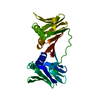
| ||||||||||||||||||
|---|---|---|---|---|---|---|---|---|---|---|---|---|---|---|---|---|---|---|---|
| 1 | 
| ||||||||||||||||||
| 2 | x 12
| ||||||||||||||||||
| Unit cell |
| ||||||||||||||||||
| Components on special symmetry positions |
| ||||||||||||||||||
| Details | The biological assembly is a trimer generated from the monomer in the asymmetric unit by applying the transformation matrix: [0 1 0] [0 0 1] [1 0 0] and [0 0 1] [1 0 0] [0 1 0] |
-Components
| #1: Protein | Mass: 27301.521 Da / Num. of mol.: 1 Source method: isolated from a genetically manipulated source Source: (gene. exp.) Archaeoglobus fulgidus (archaea) / Gene: PCN, AF0335 / Plasmid: pET11a / Species (production host): Escherichia coli / Production host:  |
|---|---|
| #2: Water | ChemComp-HOH /  Mass: 18.015 Da / Num. of mol.: 316 / Source method: isolated from a natural source / Formula: H2O Mass: 18.015 Da / Num. of mol.: 316 / Source method: isolated from a natural source / Formula: H2O |
-Experimental details
-Experiment
| Experiment | Method: X-RAY DIFFRACTION / Number of used crystals: 1 |
|---|
- Sample preparation
| Crystal grow | Temperature: 293 K / Method: vapor diffusion, hanging drop / pH: 9 Details: Na/K phosphate, pH 9.0, VAPOR DIFFUSION, HANGING DROP, temperature 293K | |||||||||||||||||||||||||||||||||||
|---|---|---|---|---|---|---|---|---|---|---|---|---|---|---|---|---|---|---|---|---|---|---|---|---|---|---|---|---|---|---|---|---|---|---|---|---|
| Crystal grow | *PLUS Temperature: 21 ℃ / pH: 8 / Method: vapor diffusion, hanging drop | |||||||||||||||||||||||||||||||||||
| Components of the solutions | *PLUS
|
-Data collection
| Diffraction | Mean temperature: 100 K |
|---|---|
| Diffraction source | Source: SYNCHROTRON / Site: APS  / Beamline: 14-BM-C / Wavelength: 1.2398 Å / Beamline: 14-BM-C / Wavelength: 1.2398 Å |
| Detector | Type: ADSC QUANTUM 4 / Detector: CCD / Date: Apr 16, 2000 |
| Radiation | Monochromator: triangle Ge(111), conical Si/Rh mirror / Protocol: SINGLE WAVELENGTH / Monochromatic (M) / Laue (L): M / Scattering type: x-ray |
| Radiation wavelength | Wavelength: 1.2398 Å / Relative weight: 1 |
| Reflection | Resolution: 1.8→30 Å / Num. all: 56483 / Num. obs: 56483 / % possible obs: 97.3 % / Observed criterion σ(F): 2 / Observed criterion σ(I): 2 / Rmerge(I) obs: 0.059 / Rsym value: 0.059 |
| Reflection shell | Resolution: 1.8→1.86 Å / Redundancy: 93.9 % / Rmerge(I) obs: 0.263 / Mean I/σ(I) obs: 5.8 / Num. unique all: 5493 / Rsym value: 0.263 / % possible all: 96.6 |
- Processing
| Software |
| ||||||||||||||||||||
|---|---|---|---|---|---|---|---|---|---|---|---|---|---|---|---|---|---|---|---|---|---|
| Refinement | Method to determine structure: MOLECULAR REPLACEMENT Starting model: Model built from 2.1 angstrom resolution data phased by a single anomalous derivative (ethyl mercury phosphate) Resolution: 1.8→30 Å / Cross valid method: THROUGHOUT / σ(F): 0 / Stereochemistry target values: Engh & Huber
| ||||||||||||||||||||
| Refinement step | Cycle: LAST / Resolution: 1.8→30 Å
| ||||||||||||||||||||
| Refine LS restraints |
| ||||||||||||||||||||
| Refine LS restraints | *PLUS
|
