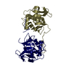
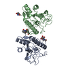
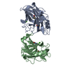
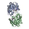
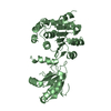
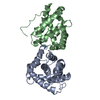
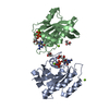
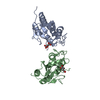
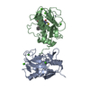
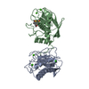
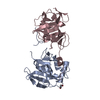
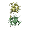
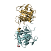
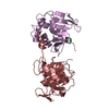
#1: Journal: J.Gen.Virol. / Year: 1998 Title: A Structural Model of Picornaviral Leader Proteinases Based on Papain and Bleomycin Hydrolase Authors: Skern, T. / Fita, I. / Guarne, A.
History
Deposition
Nov 13, 1999
Deposition site: PDBE / Processing site: PDBE
Revision 1.0
Nov 10, 2000
Provider: repository / Type: Initial release
Revision 1.1
May 8, 2011
Group: Version format compliance
Revision 1.2
Jul 13, 2011
Group: Version format compliance
Revision 1.3
Jul 24, 2019
Group: Data collection / Category: diffrn_source / Item: _diffrn_source.pdbx_synchrotron_site
Mass: 35.453 Da / Num. of mol.: 3 / Source method: obtained synthetically / Formula: Cl
Has protein modification
Y
Sequence details
THE SWISSPROT ENTRY POLG_FMDVO REFERS TO A GENOME POLYPROTEIN, AND THE PROTEASE (EC 3.4.22.-) ...THE SWISSPROT ENTRY POLG_FMDVO REFERS TO A GENOME POLYPROTEIN, AND THE PROTEASE (EC 3.4.22.-) LISTED IS FOR THE RESIDUES 1650 TO 1862. THE PROTEASE STUDIED HERE WAS FOUND IN THE RESIDUES 29 TO 195 THAT ARE LISTED IN SWISSPROT AS A NONSTRUCTURAL PROTEIN P20A.
-
Experimental details
-
Experiment
Experiment
Method: X-RAY DIFFRACTION / Number of used crystals: 1
-
Sample preparation
Crystal
Density Matthews: 3 Å3/Da / Density % sol: 58.9 %
Crystal grow
pH: 8.5 Details: 10% PEG 6000, 0.8 M MG CHLORIDE, 0.1 M TRIS/HCL PH=8.5, pH 8.50
Protocol: SINGLE WAVELENGTH / Monochromatic (M) / Laue (L): M / Scattering type: x-ray
Radiation wavelength
Wavelength: 0.8469 Å / Relative weight: 1
Reflection
Resolution: 3→34.7 Å / Num. obs: 35080 / % possible obs: 92.5 % / Redundancy: 8.4 % / Biso Wilson estimate: 63.7 Å2 / Rsym value: 0.01 / Net I/σ(I): 9.5
Reflection shell
Resolution: 3→3.05 Å / Mean I/σ(I) obs: 2.9 / Rsym value: 0.263 / % possible all: 74.9
Reflection
*PLUS
Rmerge(I) obs: 0.103
Reflection shell
*PLUS
% possible obs: 75 % / Rmerge(I) obs: 0.263
-
Processing
Software
Name
Version
Classification
CNS
0.9
refinement
DENZO
datareduction
SCALEPACK
datascaling
AMoRE
phasing
Refinement
Method to determine structure: MOLECULAR REPLACEMENT / Resolution: 3→19.99 Å / Rfactor Rfree error: 0.007 / Data cutoff high absF: 2758596.51 / Isotropic thermal model: RESTRAINED / Cross valid method: THROUGHOUT / σ(F): 0 Details: NATIVE PATTERSON MAP PRESENT A PSEUDO-ORIGEN PEAK AT POSITION (0.5,0.5,0.225). THIS PEAK CORRESPONDS A PURE TRANSLATION THAT RELATES FOUR MOLECULES OF THE A.S.U. WITH THE OTHER FOUR
In the structure databanks used in Yorodumi, some data are registered as the other names, "COVID-19 virus" and "2019-nCoV". Here are the details of the virus and the list of structure data.
Jan 31, 2019. EMDB accession codes are about to change! (news from PDBe EMDB page)
EMDB accession codes are about to change! (news from PDBe EMDB page)
The allocation of 4 digits for EMDB accession codes will soon come to an end. Whilst these codes will remain in use, new EMDB accession codes will include an additional digit and will expand incrementally as the available range of codes is exhausted. The current 4-digit format prefixed with “EMD-” (i.e. EMD-XXXX) will advance to a 5-digit format (i.e. EMD-XXXXX), and so on. It is currently estimated that the 4-digit codes will be depleted around Spring 2019, at which point the 5-digit format will come into force.
The EM Navigator/Yorodumi systems omit the EMD- prefix.
Related info.:Q: What is EMD? / ID/Accession-code notation in Yorodumi/EM Navigator
Yorodumi is a browser for structure data from EMDB, PDB, SASBDB, etc.
This page is also the successor to EM Navigator detail page, and also detail information page/front-end page for Omokage search.
The word "yorodu" (or yorozu) is an old Japanese word meaning "ten thousand". "mi" (miru) is to see.
Related info.:EMDB / PDB / SASBDB / Comparison of 3 databanks / Yorodumi Search / Aug 31, 2016. New EM Navigator & Yorodumi / Yorodumi Papers / Jmol/JSmol / Function and homology information / Changes in new EM Navigator and Yorodumi
 Movie
Movie Controller


 Open data
 Basic information
 Components
 Keywords
 Function and homology information
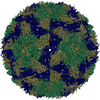
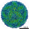 FOOT-AND-MOUTH DISEASE VIRUS
 X-RAY DIFFRACTION /
  Authors
 Citation
 Structure visualization
 Downloads & links
 Other downloads
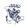
 PDBj
PDBj

 Assembly
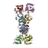


 Mass: 62.068 Da / Num. of mol.: 3 / Source method: obtained synthetically / Formula: C2H6O2
 Mass: 35.453 Da / Num. of mol.: 3 / Source method: obtained synthetically / Formula: Cl
 Sample preparation
 / Beamline: X11 / Wavelength: 0.8469
  Processing