 Movie
Movie Controller

[English] 日本語
 Yorodumi
- PDB-1khh: Crystal Structure of Guanidinoacetate Methyltransferase from Rat ... -
+ Open data

- Basic information
| Entry | Database: PDB / ID: 1khh | ||||||
|---|---|---|---|---|---|---|---|
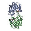
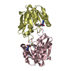
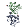
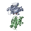
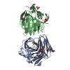
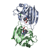
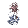
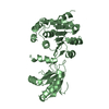
| Title | Crystal Structure of Guanidinoacetate Methyltransferase from Rat Liver: A Template Structure of Protein Arginine Methyltransferase | ||||||
 Components Components | Guanidinoacetate methyltransferase | ||||||
 Keywords Keywords | TRANSFERASE / methyltransferase / creatine biosynthesis | ||||||
| Function / homology |  Function and homology information Function and homology informationCreatine metabolism / guanidinoacetate N-methyltransferase / guanidinoacetate N-methyltransferase activity / creatine biosynthetic process / embryonic liver development / : / S-adenosylmethionine metabolic process / S-adenosylmethionine-dependent methyltransferase activity / regulation of multicellular organism growth / animal organ morphogenesis ...Creatine metabolism / guanidinoacetate N-methyltransferase / guanidinoacetate N-methyltransferase activity / creatine biosynthetic process / embryonic liver development / : / S-adenosylmethionine metabolic process / S-adenosylmethionine-dependent methyltransferase activity / regulation of multicellular organism growth / animal organ morphogenesis / methylation / spermatogenesis / identical protein binding / nucleus / cytoplasm Similarity search - Function | ||||||
| Biological species |  | ||||||
| Method |  X-RAY DIFFRACTION / SIR / Resolution: 2.5 Å X-RAY DIFFRACTION / SIR / Resolution: 2.5 Å | ||||||
 Authors Authors | Takusagawa, F. / Komoto, J. | ||||||
 Citation Citation | Journal: J.Mol.Biol. / Year: 2002 Title: Crystal structure of guanidinoacetate methyltransferase from rat liver: a model structure of protein arginine methyltransferase. Authors: Komoto, J. / Huang, Y. / Takata, Y. / Yamada, T. / Konishi, K. / Ogawa, H. / Gomi, T. / Fujioka, M. / Takusagawa, F. | ||||||
| History |
| ||||||
| Remark 300 | BIOMOLECULE: 1 THIS ENTRY CONTAINS THE CRYSTALLOGRAPHIC ASYMMETRIC UNIT WHICH CONSISTS OF 2 ... BIOMOLECULE: 1 THIS ENTRY CONTAINS THE CRYSTALLOGRAPHIC ASYMMETRIC UNIT WHICH CONSISTS OF 2 CHAIN(S). SEE REMARK 350 FOR INFORMATION ON GENERATING THE BIOLOGICAL MOLECULE(S). THE TRUNCATED ENZYME FORMS A HOMODIMER. | ||||||
| Remark 999 | SEQUENCE According to the authors, the correct amino acid residue is Val (GTG) at position 119. ...SEQUENCE According to the authors, the correct amino acid residue is Val (GTG) at position 119. Therefore, the sequence in the PDB file is the updated and correct one. |
- Structure visualization
| Structure viewer | Molecule: MolmilJmol/JSmol |
|---|
- Downloads & links
-Download
| PDBx/mmCIF format | 1khh.cif.gz | 93.6 KB | Display | PDBx/mmCIF format |
|---|---|---|---|---|
| PDB format | pdb1khh.ent.gz | 70.1 KB | Display | PDB format |
| PDBx/mmJSON format | 1khh.json.gz | Tree view | PDBx/mmJSON format | |
| Others |  Other downloads Other downloads |
-Validation report
| Arichive directory | https://data.pdbj.org/pub/pdb/validation_reports/kh/1khhftp://data.pdbj.org/pub/pdb/validation_reports/kh/1khh | HTTPS FTP |
|---|
-Related structure data
| Similar structure data |
|---|


-Links
 PDBj
PDBj- Assembly
| Deposited unit | 
| ||||||||
|---|---|---|---|---|---|---|---|---|---|
| 1 |
| ||||||||
| Unit cell |
| ||||||||
| Details | The biological assembly is a monomer. However, the N-terminal truncated enzyme forms a homodimer. |
-Components
| #1: Protein | Mass: 22557.988 Da / Num. of mol.: 2 / Fragment: N-terminal truncation Source method: isolated from a genetically manipulated source Source: (gene. exp.)  References: UniProt: P10868, guanidinoacetate N-methyltransferase #2: Chemical |   Type: L-peptide linking / Mass: 384.411 Da / Num. of mol.: 2 / Source method: obtained synthetically / Formula: C14H20N6O5S Type: L-peptide linking / Mass: 384.411 Da / Num. of mol.: 2 / Source method: obtained synthetically / Formula: C14H20N6O5S#3: Water | ChemComp-HOH / |  Mass: 18.015 Da / Num. of mol.: 162 / Source method: isolated from a natural source / Formula: H2O Mass: 18.015 Da / Num. of mol.: 162 / Source method: isolated from a natural source / Formula: H2O |
|---|
-Experimental details
-Experiment
| Experiment | Method: X-RAY DIFFRACTION / Number of used crystals: 1 |
|---|
- Sample preparation
| Crystal | Density Matthews: 2.55 Å3/Da / Density % sol: 51.8 % | ||||||||||||||||||||||||||||||||||||||||||
|---|---|---|---|---|---|---|---|---|---|---|---|---|---|---|---|---|---|---|---|---|---|---|---|---|---|---|---|---|---|---|---|---|---|---|---|---|---|---|---|---|---|---|---|
| Crystal grow | Temperature: 277 K / Method: vapor diffusion, hanging drop / pH: 6.5 Details: PEG 4000, pH 6.5, VAPOR DIFFUSION, HANGING DROP, temperature 277K | ||||||||||||||||||||||||||||||||||||||||||
| Crystal grow | *PLUS Temperature: 4 ℃ | ||||||||||||||||||||||||||||||||||||||||||
| Components of the solutions | *PLUS
|
-Data collection
| Diffraction | Mean temperature: 93 K |
|---|---|
| Diffraction source | Source: ROTATING ANODE / Type: RIGAKU RU200 / Wavelength: 1.5418 Å |
| Detector | Type: RIGAKU RAXIS IIC / Detector: IMAGE PLATE / Date: May 1, 2001 / Details: Confocal optics |
| Radiation | Protocol: SINGLE WAVELENGTH / Monochromatic (M) / Laue (L): M / Scattering type: x-ray |
| Radiation wavelength | Wavelength: 1.5418 Å / Relative weight: 1 |
| Reflection | Resolution: 2.5→34.5 Å / Num. all: 109295 / Num. obs: 109295 / % possible obs: 98 % / Observed criterion σ(F): 0 / Observed criterion σ(I): 0 / Redundancy: 6.7 % / Biso Wilson estimate: 14.7 Å2 / Rmerge(I) obs: 0.047 / Rsym value: 0.047 / Net I/σ(I): 11.4 |
| Reflection shell | Resolution: 2.5→2.58 Å / Redundancy: 3.1 % / Rmerge(I) obs: 0.139 / Mean I/σ(I) obs: 5 / Num. unique all: 1332 / Rsym value: 0.139 / % possible all: 83.4 |
| Reflection | *PLUS Highest resolution: 2.5 Å / Num. obs: 15472 / % possible obs: 98 % / Num. measured all: 109295 |
- Processing
| Software |
| |||||||||||||||||||||||||
|---|---|---|---|---|---|---|---|---|---|---|---|---|---|---|---|---|---|---|---|---|---|---|---|---|---|---|
| Refinement | Method to determine structure: SIR / Resolution: 2.5→8 Å / σ(F): 2 / Stereochemistry target values: Engh & Huber Details: The structure was solved by SIR using Gd-derivative. Residues 38-42 are disordered.
| |||||||||||||||||||||||||
| Refine analyze | Luzzati coordinate error obs: 0.03 Å | |||||||||||||||||||||||||
| Refinement step | Cycle: LAST / Resolution: 2.5→8 Å
| |||||||||||||||||||||||||
| Refine LS restraints |
| |||||||||||||||||||||||||
| LS refinement shell | Refine-ID: X-RAY DIFFRACTION
| |||||||||||||||||||||||||
| Software | *PLUS Name: X-PLOR / Version: 3.843 / Classification: refinement | |||||||||||||||||||||||||
| Refinement | *PLUS Highest resolution: 2.5 Å / Lowest resolution: 8 Å / σ(F): 2 / Num. reflection Rfree: 1481 / Rfactor obs: 0.214 | |||||||||||||||||||||||||
| Solvent computation | *PLUS | |||||||||||||||||||||||||
| Displacement parameters | *PLUS | |||||||||||||||||||||||||
| Refine LS restraints | *PLUS
| |||||||||||||||||||||||||
| LS refinement shell | *PLUS Highest resolution: 2.5 Å / Rfactor Rfree: 0.376 / Rfactor Rwork: 0.288 / Rfactor obs: 0.288 |
