 Movie
Movie Controller

[English] 日本語
 Yorodumi
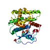
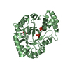
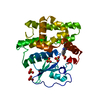
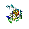
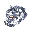
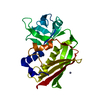
- PDB-1qls: S100C (S100A11),OR CALGIZZARIN, IN COMPLEX WITH ANNEXIN I N-TERMINUS -
+ Open data

- Basic information
| Entry | Database: PDB / ID: 1qls | ||||||
|---|---|---|---|---|---|---|---|
| Title | S100C (S100A11),OR CALGIZZARIN, IN COMPLEX WITH ANNEXIN I N-TERMINUS | ||||||
 Components Components |
| ||||||
 Keywords Keywords | METAL-BINDING PROTEIN/INHIBITOR / S100 FAMILY / EF-HAND PROTEIN / COMPLEX (LIGAND-ANNEXIN) / LIGAND OF ANNEXIN II / CALCIUM/PHOSPHOLIPID BINDING PROTEIN / METAL-BINDING PROTEIN-INHIBITOR complex | ||||||
| Function / homology |  Function and homology information Function and homology informationregulation of interleukin-1 production / myoblast migration involved in skeletal muscle regeneration / granulocyte chemotaxis / regulation of leukocyte migration / positive regulation of T-helper 1 cell differentiation / phospholipase A2 inhibitor activity / positive regulation of vesicle fusion / regulation of hormone secretion / neutrophil clearance / positive regulation of neutrophil apoptotic process ...regulation of interleukin-1 production / myoblast migration involved in skeletal muscle regeneration / granulocyte chemotaxis / regulation of leukocyte migration / positive regulation of T-helper 1 cell differentiation / phospholipase A2 inhibitor activity / positive regulation of vesicle fusion / regulation of hormone secretion / neutrophil clearance / positive regulation of neutrophil apoptotic process / peptide cross-linking / cadherin binding involved in cell-cell adhesion / cornified envelope / neutrophil activation / negative regulation of interleukin-8 production / neutrophil homeostasis / calcium-dependent phospholipid binding / negative regulation of T-helper 2 cell differentiation / Neutrophil degranulation / S100 protein binding / Formyl peptide receptors bind formyl peptides and many other ligands / vesicle membrane / positive regulation of cell migration involved in sprouting angiogenesis / alpha-beta T cell differentiation / negative regulation of exocytosis / arachidonate secretion / cellular response to glucocorticoid stimulus / motile cilium / positive regulation of wound healing / phosphatidylserine binding / monocyte chemotaxis / lateral plasma membrane / cellular response to vascular endothelial growth factor stimulus / Smooth Muscle Contraction / positive regulation of G1/S transition of mitotic cell cycle / phagocytosis / G protein-coupled receptor signaling pathway, coupled to cyclic nucleotide second messenger / keratinocyte differentiation / phagocytic cup / Developmental Lineage of Pancreatic Ductal Cells / positive regulation of interleukin-2 production / positive regulation of T cell proliferation / adherens junction / sarcolemma / phospholipid binding / extracellular matrix / calcium-dependent protein binding / regulation of cell shape / regulation of cell population proliferation / actin cytoskeleton organization / regulation of inflammatory response / early endosome membrane / Interleukin-4 and Interleukin-13 signaling / G alpha (i) signalling events / basolateral plasma membrane / vesicle / G alpha (q) signalling events / adaptive immune response / cell surface receptor signaling pathway / apical plasma membrane / endosome / inflammatory response / signaling receptor binding / innate immune response / focal adhesion / calcium ion binding / lipid binding / negative regulation of apoptotic process / cell surface / signal transduction / extracellular space / extracellular exosome / extracellular region / nucleoplasm / nucleus / plasma membrane / cytoplasm / cytosol Similarity search - Function | ||||||
| Biological species |   HOMO SAPIENS (human) HOMO SAPIENS (human) | ||||||
| Method |  X-RAY DIFFRACTION / SYNCHROTRON / MOLECULAR REPLACEMENT / Resolution: 2.3 Å X-RAY DIFFRACTION / SYNCHROTRON / MOLECULAR REPLACEMENT / Resolution: 2.3 Å | ||||||
 Authors Authors | Rety, S. / Sopkova, J. / Renouard, M. / Osterloh, D. / Gerke, V. / Russo-Marie, F. / Lewit-Bentley, A. | ||||||
 Citation Citation | Journal: Structure / Year: 2000 Title: Structural Basis of the Ca2+ Dependent Association between S100C (S100A11) and its Target, the N-Terminal Part of Annexin I Authors: Rety, S. / Osterloh, D. / Arie, J.P. / Tabaries, S. / Seeman, J. / Russo-Marie, F. / Gerke, V. / Lewit-Bentley, A. | ||||||
| History |
| ||||||
| Remark 650 | HELIX DETERMINATION METHOD: AUTHOR PROVIDED. | ||||||
| Remark 700 | SHEET DETERMINATION METHOD: AUTHOR PROVIDED. |
- Structure visualization
| Structure viewer | Molecule: MolmilJmol/JSmol |
|---|
- Downloads & links
-Download
| PDBx/mmCIF format | 1qls.cif.gz | 36.3 KB | Display | PDBx/mmCIF format |
|---|---|---|---|---|
| PDB format | pdb1qls.ent.gz | 24 KB | Display | PDB format |
| PDBx/mmJSON format | 1qls.json.gz | Tree view | PDBx/mmJSON format | |
| Others |  Other downloads Other downloads |
-Validation report
| Arichive directory | https://data.pdbj.org/pub/pdb/validation_reports/ql/1qlsftp://data.pdbj.org/pub/pdb/validation_reports/ql/1qls | HTTPS FTP |
|---|
-Related structure data
| Related structure data |  1bt6S S: Starting model for refinement |
|---|---|
| Similar structure data |


-Links
 PDBj
PDBj


- Assembly
| Deposited unit | 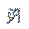
| ||||||||
|---|---|---|---|---|---|---|---|---|---|
| 1 | 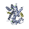
| ||||||||
| Unit cell |
| ||||||||
| Details | BIOLOGICAL_UNIT: DIMERTHE DIMER IS COVALENT IN THE CRYSTAL, LINKED BY ADISULPHIDE AT CYS11. UNDER REDUCING CONDITIONS, THOUGHTHE DISULPHIDE WOULD BE REDUCED, THE DIMER SHOULD REMAININTACT.FOR THE HOMO-ASSEMBLY DESCRIBED BY REMARK 350THE DIFFERENCE IN ACCESSIBLE SURFACE AREA PERCHAIN BETWEEN THE ISOLATED CHAIN AND THAT FOR |
-Components
| #1: Protein | Mass: 11194.829 Da / Num. of mol.: 1 Source method: isolated from a genetically manipulated source Source: (gene. exp.)  | ||||||
|---|---|---|---|---|---|---|---|
| #2: Protein/peptide | Mass: 1278.541 Da / Num. of mol.: 1 / Fragment: N-TERMINAL / Source method: obtained synthetically / Details: N-ACETYLATED ON N-TERMINUS / Source: (synth.) HOMO SAPIENS (human) / References: UniProt: P04083 | ||||||
| #3: Chemical |   Mass: 40.078 Da / Num. of mol.: 2 / Source method: obtained synthetically / Formula: Ca Mass: 40.078 Da / Num. of mol.: 2 / Source method: obtained synthetically / Formula: Ca#4: Water | ChemComp-HOH / |  Mass: 18.015 Da / Num. of mol.: 23 / Source method: isolated from a natural source / Formula: H2O Mass: 18.015 Da / Num. of mol.: 23 / Source method: isolated from a natural source / Formula: H2OHas protein modification | Y | Sequence details | SYNTHETIC ANNEXIN I N-TERMINAL SEQUENCE | |
-Experimental details
-Experiment
| Experiment | Method: X-RAY DIFFRACTION / Number of used crystals: 1 |
|---|
- Sample preparation
| Crystal | Density Matthews: 3.9 Å3/Da / Density % sol: 68.2 % | |||||||||||||||||||||||||
|---|---|---|---|---|---|---|---|---|---|---|---|---|---|---|---|---|---|---|---|---|---|---|---|---|---|---|
| Crystal grow | Method: vapor diffusion / pH: 8.5 Details: 20 MG/ML PROTEIN WERE CRYSTALLIZED BY VAPOR DIFFUSION AGAINST 10% PEG 4000, 20% PEG 4000, 10% 2-PROPANOL, 100MM HEPES, PH=8.5, pH 8.50 | |||||||||||||||||||||||||
| Crystal grow | *PLUS Method: vapor diffusion, hanging dropDetails: drop consists of 1:1 mixture of well and protein solutions | |||||||||||||||||||||||||
| Components of the solutions | *PLUS
|
-Data collection
| Diffraction | Mean temperature: 280 K |
|---|---|
| Diffraction source | Source: SYNCHROTRON / Site: LURE  / Beamline: D41A / Wavelength: 1.37 / Beamline: D41A / Wavelength: 1.37 |
| Detector | Type: MARRESEARCH / Detector: IMAGE PLATE / Date: Jul 2, 1998 / Details: FOCUSSING MONOCHROMATOR |
| Radiation | Monochromator: GE(111) / Protocol: SINGLE WAVELENGTH / Monochromatic (M) / Laue (L): M / Scattering type: x-ray |
| Radiation wavelength | Wavelength: 1.37 Å / Relative weight: 1 |
| Reflection | Resolution: 2.3→69 Å / Num. obs: 9310 / % possible obs: 99.7 % / Observed criterion σ(I): 0 / Redundancy: 6.6 % / Rmerge(I) obs: 0.08 / Rsym value: 0.08 / Net I/σ(I): 20.1 |
| Reflection | *PLUS Lowest resolution: 20 Å / Num. measured all: 61772 |
- Processing
| Software |
| ||||||||||||||||||||||||||||||||||||||||||||||||||||||||||||||||||||||||||||||||||||
|---|---|---|---|---|---|---|---|---|---|---|---|---|---|---|---|---|---|---|---|---|---|---|---|---|---|---|---|---|---|---|---|---|---|---|---|---|---|---|---|---|---|---|---|---|---|---|---|---|---|---|---|---|---|---|---|---|---|---|---|---|---|---|---|---|---|---|---|---|---|---|---|---|---|---|---|---|---|---|---|---|---|---|---|---|---|
| Refinement | Method to determine structure: MOLECULAR REPLACEMENT Starting model: 1BT6 Resolution: 2.3→20 Å / SU B: 4.869 / SU ML: 0.1216 / Cross valid method: THROUGHOUT / σ(F): 0 / ESU R: 0.217 / ESU R Free: 0.207
| ||||||||||||||||||||||||||||||||||||||||||||||||||||||||||||||||||||||||||||||||||||
| Displacement parameters | Biso mean: 200.109 Å2 | ||||||||||||||||||||||||||||||||||||||||||||||||||||||||||||||||||||||||||||||||||||
| Refinement step | Cycle: LAST / Resolution: 2.3→20 Å
| ||||||||||||||||||||||||||||||||||||||||||||||||||||||||||||||||||||||||||||||||||||
| Refine LS restraints |
| ||||||||||||||||||||||||||||||||||||||||||||||||||||||||||||||||||||||||||||||||||||
| Software | *PLUS Name: REFMAC / Classification: refinement | ||||||||||||||||||||||||||||||||||||||||||||||||||||||||||||||||||||||||||||||||||||
| Refine LS restraints | *PLUS
|
