 Movie
Movie Controller


+ Open data

- Basic information
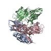
| Entry | Database: PDB / ID: 1a4p | ||||||
|---|---|---|---|---|---|---|---|
| Title | P11 (S100A10), LIGAND OF ANNEXIN II | ||||||
 Components Components | S100A10 | ||||||
 Keywords Keywords | CALCIUM/PHOSPHOLIPID BINDING PROTEIN / S100 FAMILY / EF-HAND PROTEIN / LIGAND OF ANNEXIN II / CALCIUM-PHOSPHOLIPID BINDING PROTEIN complex | ||||||
| Function / homology |  Function and homology information Function and homology informationAnxA2-p11 complex / membrane raft assembly / positive regulation of plasma membrane repair / positive regulation of plasminogen activation / vesicle budding from membrane / plasma membrane protein complex / Dissolution of Fibrin Clot / positive regulation of exocytosis / positive regulation of GTPase activity / positive regulation of focal adhesion assembly ...AnxA2-p11 complex / membrane raft assembly / positive regulation of plasma membrane repair / positive regulation of plasminogen activation / vesicle budding from membrane / plasma membrane protein complex / Dissolution of Fibrin Clot / positive regulation of exocytosis / positive regulation of GTPase activity / positive regulation of focal adhesion assembly / regulation of neurogenesis / positive regulation of stress fiber assembly / positive regulation of substrate adhesion-dependent cell spreading / protein localization to plasma membrane / mRNA transcription by RNA polymerase II / RNA polymerase II transcription regulator complex / nuclear matrix / calcium-dependent protein binding / extracellular matrix / transmembrane transporter binding / calcium ion binding / cell surface / protein homodimerization activity / positive regulation of transcription by RNA polymerase II / extracellular exosome / extracellular region / plasma membrane / cytoplasm Similarity search - Function | ||||||
| Biological species |  Homo sapiens (human) Homo sapiens (human) | ||||||
| Method |  X-RAY DIFFRACTION / SYNCHROTRON / MIR / Resolution: 2.25 Å X-RAY DIFFRACTION / SYNCHROTRON / MIR / Resolution: 2.25 Å | ||||||
 Authors Authors | Rety, S. / Sopkova, J. / Renouard, M. / Osterloh, D. / Gerke, V. / Russo-Marie, F. / Lewit-Bentley, A. | ||||||
 Citation Citation | Journal: Nat.Struct.Biol. / Year: 1999 Title: The crystal structure of a complex of p11 with the annexin II N-terminal peptide. Authors: Rety, S. / Sopkova, J. / Renouard, M. / Osterloh, D. / Gerke, V. / Tabaries, S. / Russo-Marie, F. / Lewit-Bentley, A. | ||||||
| History |
|
- Structure visualization
| Structure viewer | Molecule: MolmilJmol/JSmol |
|---|
- Downloads & links
-Download
| PDBx/mmCIF format | 1a4p.cif.gz | 49.4 KB | Display | PDBx/mmCIF format |
|---|---|---|---|---|
| PDB format | pdb1a4p.ent.gz | 36.5 KB | Display | PDB format |
| PDBx/mmJSON format | 1a4p.json.gz | Tree view | PDBx/mmJSON format | |
| Others |  Other downloads Other downloads |
-Validation report
| Arichive directory | https://data.pdbj.org/pub/pdb/validation_reports/a4/1a4pftp://data.pdbj.org/pub/pdb/validation_reports/a4/1a4p | HTTPS FTP |
|---|
-Related structure data


-Links
 PDBj
PDBj- Assembly
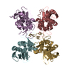
| Deposited unit | 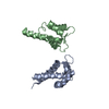
| ||||||||
|---|---|---|---|---|---|---|---|---|---|
| 1 | 
| ||||||||
| 2 | 
| ||||||||
| Unit cell |
| ||||||||
| Noncrystallographic symmetry (NCS) | NCS oper: (Code: given Matrix: (-0.85907, -0.47311, -0.19537), Vector: |
-Components
| #1: Protein | Mass: 11088.940 Da / Num. of mol.: 2 Source method: isolated from a genetically manipulated source Source: (gene. exp.) Homo sapiens (human) / Plasmid: PET23A / Species (production host): Escherichia coli / Production host:  #2: Water | ChemComp-HOH / |  Mass: 18.015 Da / Num. of mol.: 63 / Source method: isolated from a natural source / Formula: H2O Mass: 18.015 Da / Num. of mol.: 63 / Source method: isolated from a natural source / Formula: H2OHas protein modification | Y | |
|---|
-Experimental details
-Experiment
| Experiment | Method: X-RAY DIFFRACTION / Number of used crystals: 1 |
|---|
- Sample preparation
| Crystal | Density Matthews: 2.53 Å3/Da / Density % sol: 51 % | ||||||||||||||||||||||||||||||||||||||||||||||||
|---|---|---|---|---|---|---|---|---|---|---|---|---|---|---|---|---|---|---|---|---|---|---|---|---|---|---|---|---|---|---|---|---|---|---|---|---|---|---|---|---|---|---|---|---|---|---|---|---|---|
| Crystal grow | Method: vapor diffusion / pH: 7.5 Details: 15 MG/ML PROTEIN WERE CRYSTALLIZED BY VAPOR DIFFUSION AGAINST 20% PEG 4000, 10% 2-PROPANOL, 100MM HEPES, PH=7.5, vapor diffusion | ||||||||||||||||||||||||||||||||||||||||||||||||
| Crystal grow | *PLUS Method: vapor diffusion, hanging drop / Details: drop:reservoir=1:1 | ||||||||||||||||||||||||||||||||||||||||||||||||
| Components of the solutions | *PLUS
|
-Data collection
| Diffraction | Mean temperature: 280 K |
|---|---|
| Diffraction source | Source: SYNCHROTRON / Site: LURE  / Beamline: D41A / Wavelength: 1.37 / Beamline: D41A / Wavelength: 1.37 |
| Detector | Type: MARRESEARCH / Detector: IMAGE PLATE / Date: Jul 18, 1997 / Details: FOCUSSING MONOCHROMATOR |
| Radiation | Monochromator: GE(111) / Monochromatic (M) / Laue (L): M / Scattering type: x-ray |
| Radiation wavelength | Wavelength: 1.37 Å / Relative weight: 1 |
| Reflection | Resolution: 2.25→62 Å / Num. obs: 10769 / % possible obs: 98.4 % / Observed criterion σ(I): 0 / Redundancy: 6.15 % / Rsym value: 0.047 / Net I/σ(I): 14.9 |
| Reflection shell | Resolution: 2.25→2.33 Å / Redundancy: 3.5 % / Mean I/σ(I) obs: 3.5 / Rsym value: 0.168 / % possible all: 97.1 |
| Reflection | *PLUS Num. obs: 10957 / Num. measured all: 67368 / Rmerge(I) obs: 0.032 |
| Reflection shell | *PLUS % possible obs: 97.1 % / Rmerge(I) obs: 0.168 |
- Processing
| Software |
| ||||||||||||||||||||||||||||||||||||||||||||||||||||||||||||||||||||||||||||||||||||
|---|---|---|---|---|---|---|---|---|---|---|---|---|---|---|---|---|---|---|---|---|---|---|---|---|---|---|---|---|---|---|---|---|---|---|---|---|---|---|---|---|---|---|---|---|---|---|---|---|---|---|---|---|---|---|---|---|---|---|---|---|---|---|---|---|---|---|---|---|---|---|---|---|---|---|---|---|---|---|---|---|---|---|---|---|---|
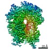
| Refinement | Method to determine structure: MIR / Resolution: 2.25→20 Å / Cross valid method: THROUGHOUT / σ(F): 0
| ||||||||||||||||||||||||||||||||||||||||||||||||||||||||||||||||||||||||||||||||||||
| Displacement parameters | Biso mean: 36.1 Å2 | ||||||||||||||||||||||||||||||||||||||||||||||||||||||||||||||||||||||||||||||||||||
| Refine analyze | Luzzati coordinate error obs: 0.3 Å / Luzzati d res low obs: 20 Å | ||||||||||||||||||||||||||||||||||||||||||||||||||||||||||||||||||||||||||||||||||||
| Refinement step | Cycle: LAST / Resolution: 2.25→20 Å
| ||||||||||||||||||||||||||||||||||||||||||||||||||||||||||||||||||||||||||||||||||||
| Refine LS restraints |
| ||||||||||||||||||||||||||||||||||||||||||||||||||||||||||||||||||||||||||||||||||||
| Software | *PLUS Name: REFMAC / Classification: refinement | ||||||||||||||||||||||||||||||||||||||||||||||||||||||||||||||||||||||||||||||||||||
| Refinement | *PLUS Rfactor all: 0.246 / Rfactor obs: 0.227 | ||||||||||||||||||||||||||||||||||||||||||||||||||||||||||||||||||||||||||||||||||||
| Solvent computation | *PLUS | ||||||||||||||||||||||||||||||||||||||||||||||||||||||||||||||||||||||||||||||||||||
| Displacement parameters | *PLUS |
