 Movie
Movie Controller

[English] 日本語
 Yorodumi
- PDB-1bt6: P11 (S100A10), LIGAND OF ANNEXIN II IN COMPLEX WITH ANNEXIN II N-... -
+ Open data

- Basic information
| Entry | Database: PDB / ID: 1bt6 | ||||||
|---|---|---|---|---|---|---|---|
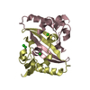
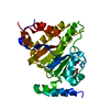
| Title | P11 (S100A10), LIGAND OF ANNEXIN II IN COMPLEX WITH ANNEXIN II N-TERMINUS | ||||||
 Components Components |
| ||||||
 Keywords Keywords | COMPLEX (LIGAND/ANNEXIN) / S100 FAMILY / EF-HAND PROTEIN / COMPLEX (LIGAND-ANNEXIN) / LIGAND OF ANNEXIN II / CALCIUM/PHOSPHOLIPID BINDING PROTEIN / COMPLEX (LIGAND-ANNEXIN) complex | ||||||
| Function / homology |  Function and homology information Function and homology informationvoltage-gated calcium channel activity involved in regulation of cytosolic calcium levels / Dissolution of Fibrin Clot / phospholipase inhibitor activity / Turbulent (oscillatory, disturbed) flow shear stress activates signaling by PIEZO1 and integrins in endothelial cells / AnxA2-p11 complex / membrane raft assembly / growth plate cartilage development / endocardial cell differentiation / positive regulation of plasma membrane repair / positive regulation of plasminogen activation ...voltage-gated calcium channel activity involved in regulation of cytosolic calcium levels / Dissolution of Fibrin Clot / phospholipase inhibitor activity / Turbulent (oscillatory, disturbed) flow shear stress activates signaling by PIEZO1 and integrins in endothelial cells / AnxA2-p11 complex / membrane raft assembly / growth plate cartilage development / endocardial cell differentiation / positive regulation of plasma membrane repair / positive regulation of plasminogen activation / positive regulation of chondrocyte differentiation / vesicle budding from membrane / calcium-dependent phospholipid binding / positive regulation of calcium ion transport / plasma membrane protein complex / Dissolution of Fibrin Clot / virion binding / vesicle membrane / phosphatidylserine binding / bone mineralization / positive regulation of transforming growth factor beta receptor signaling pathway / positive regulation of exocytosis / basement membrane / positive regulation of focal adhesion assembly / positive regulation of GTPase activity / regulation of neurogenesis / calcium ion homeostasis / cytoskeletal protein binding / positive regulation of stress fiber assembly / positive regulation of substrate adhesion-dependent cell spreading / calcium channel complex / Neutrophil degranulation / protein localization to plasma membrane / mRNA transcription by RNA polymerase II / RNA polymerase II transcription regulator complex / calcium channel activity / nuclear matrix / calcium-dependent protein binding / extracellular matrix / vesicle / transmembrane transporter binding / cell adhesion / calcium ion binding / cell surface / protein homodimerization activity / positive regulation of transcription by RNA polymerase II / protein-containing complex / extracellular exosome / extracellular region / nucleus / plasma membrane / cytoplasm Similarity search - Function | ||||||
| Biological species |  Homo sapiens (human) Homo sapiens (human) | ||||||
| Method |  X-RAY DIFFRACTION / SYNCHROTRON / MOLECULAR REPLACEMENT / Resolution: 2.4 Å X-RAY DIFFRACTION / SYNCHROTRON / MOLECULAR REPLACEMENT / Resolution: 2.4 Å | ||||||
 Authors Authors | Rety, S. / Sopkova, J. / Renouard, M. / Osterloh, D. / Gerke, V. / Russo-Marie, F. / Lewit-Bentley, A. | ||||||
 Citation Citation | Journal: Nat.Struct.Biol. / Year: 1999 Title: The crystal structure of a complex of p11 with the annexin II N-terminal peptide. Authors: Rety, S. / Sopkova, J. / Renouard, M. / Osterloh, D. / Gerke, V. / Tabaries, S. / Russo-Marie, F. / Lewit-Bentley, A. | ||||||
| History |
|
- Structure visualization
| Structure viewer | Molecule: MolmilJmol/JSmol |
|---|
- Downloads & links
-Download
| PDBx/mmCIF format | 1bt6.cif.gz | 52.4 KB | Display | PDBx/mmCIF format |
|---|---|---|---|---|
| PDB format | pdb1bt6.ent.gz | 39.2 KB | Display | PDB format |
| PDBx/mmJSON format | 1bt6.json.gz | Tree view | PDBx/mmJSON format | |
| Others |  Other downloads Other downloads |
-Validation report
| Arichive directory | https://data.pdbj.org/pub/pdb/validation_reports/bt/1bt6ftp://data.pdbj.org/pub/pdb/validation_reports/bt/1bt6 | HTTPS FTP |
|---|
-Related structure data
| Related structure data |  1a4pSC S: Starting model for refinement C: citing same article ( |
|---|---|
| Similar structure data |


-Links
 PDBj
PDBj- Assembly
| Deposited unit | 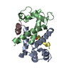
| ||||||||||||
|---|---|---|---|---|---|---|---|---|---|---|---|---|---|
| 1 |
| ||||||||||||
| 2 | 
| ||||||||||||
| Unit cell |
| ||||||||||||
| Noncrystallographic symmetry (NCS) | NCS oper:
|
-Components
| #1: Protein | Mass: 11088.940 Da / Num. of mol.: 2 Source method: isolated from a genetically manipulated source Source: (gene. exp.) Homo sapiens (human) / Plasmid: PET23A / Species (production host): Escherichia coli / Production host:  #2: Protein/peptide | Mass: 1483.706 Da / Num. of mol.: 2 / Fragment: N-TERMINAL Source method: isolated from a genetically manipulated source References: UniProt: P17785 #3: Water | ChemComp-HOH / |  Mass: 18.015 Da / Num. of mol.: 22 / Source method: isolated from a natural source / Formula: H2O Mass: 18.015 Da / Num. of mol.: 22 / Source method: isolated from a natural source / Formula: H2OHas protein modification | Y | |
|---|
-Experimental details
-Experiment
| Experiment | Method: X-RAY DIFFRACTION / Number of used crystals: 3 |
|---|
- Sample preparation
| Crystal | Density Matthews: 2.77 Å3/Da / Density % sol: 56 % | |||||||||||||||||||||||||||||||||||
|---|---|---|---|---|---|---|---|---|---|---|---|---|---|---|---|---|---|---|---|---|---|---|---|---|---|---|---|---|---|---|---|---|---|---|---|---|
| Crystal grow | pH: 7.5 / Details: pH 7.5 | |||||||||||||||||||||||||||||||||||
| Crystal grow | *PLUS Method: unknown | |||||||||||||||||||||||||||||||||||
| Components of the solutions | *PLUS
|
-Data collection
| Diffraction | Mean temperature: 280 K |
|---|---|
| Diffraction source | Source: SYNCHROTRON / Site: LURE  / Beamline: DW32 / Wavelength: 0.97 / Beamline: DW32 / Wavelength: 0.97 |
| Detector | Type: MARRESEARCH / Detector: IMAGE PLATE / Date: Apr 25, 1998 / Details: FOCUSSING MONOCHROMATOR AND MONOLAYER |
| Radiation | Monochromator: GE(111) / Monochromatic (M) / Laue (L): M / Scattering type: x-ray |
| Radiation wavelength | Wavelength: 0.97 Å / Relative weight: 1 |
| Reflection | Resolution: 2.4→62 Å / Num. obs: 9774 / % possible obs: 96.8 % / Observed criterion σ(I): 0 / Redundancy: 6.25 % / Rmerge(I) obs: 0.06 / Rsym value: 0.06 / Net I/σ(I): 20.6 |
| Reflection shell | Resolution: 2.4→2.5 Å / Redundancy: 2.5 % / Mean I/σ(I) obs: 1.96 / Rsym value: 0.61 |
| Reflection | *PLUS Num. measured all: 29481 |
| Reflection shell | *PLUS Rmerge(I) obs: 0.61 |
- Processing
| Software |
| ||||||||||||||||||||||||||||||||||||||||||||||||||||||||||||||||||||||||||||||||||||
|---|---|---|---|---|---|---|---|---|---|---|---|---|---|---|---|---|---|---|---|---|---|---|---|---|---|---|---|---|---|---|---|---|---|---|---|---|---|---|---|---|---|---|---|---|---|---|---|---|---|---|---|---|---|---|---|---|---|---|---|---|---|---|---|---|---|---|---|---|---|---|---|---|---|---|---|---|---|---|---|---|---|---|---|---|---|
| Refinement | Method to determine structure: MOLECULAR REPLACEMENT Starting model: 1A4P Resolution: 2.4→20 Å / Cross valid method: THROUGHOUT / σ(F): 0
| ||||||||||||||||||||||||||||||||||||||||||||||||||||||||||||||||||||||||||||||||||||
| Displacement parameters | Biso mean: 200.1 Å2 | ||||||||||||||||||||||||||||||||||||||||||||||||||||||||||||||||||||||||||||||||||||
| Refinement step | Cycle: LAST / Resolution: 2.4→20 Å
| ||||||||||||||||||||||||||||||||||||||||||||||||||||||||||||||||||||||||||||||||||||
| Refine LS restraints |
| ||||||||||||||||||||||||||||||||||||||||||||||||||||||||||||||||||||||||||||||||||||
| Software | *PLUS Name: REFMAC / Classification: refinement | ||||||||||||||||||||||||||||||||||||||||||||||||||||||||||||||||||||||||||||||||||||
| Refine LS restraints | *PLUS
|
