 Movie
Movie Controller

[English] 日本語
 Yorodumi
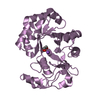
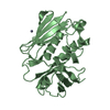
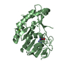
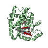
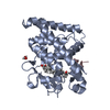
- PDB-1cqv: CRYSTAL STRUCTURE OF STAPHYLOCOCCAL ENTEROTOXIN C2 AT 100K CRYSTA... -
+ Open data

- Basic information
| Entry | Database: PDB / ID: 1cqv | ||||||
|---|---|---|---|---|---|---|---|
| Title | CRYSTAL STRUCTURE OF STAPHYLOCOCCAL ENTEROTOXIN C2 AT 100K CRYSTALLIZED AT PH 5.0 | ||||||
 Components Components | PROTEIN (STAPHYLOCOCCAL ENTEROTOXIN C2) | ||||||
 Keywords Keywords | TOXIN / ENTEROTOXIN / SUPERANTIGEN / ZINC BINDING / IMMUNE SYSTEM | ||||||
| Function / homology |  Function and homology information Function and homology information | ||||||
| Biological species |   Staphylococcus aureus (bacteria) Staphylococcus aureus (bacteria) | ||||||
| Method |  X-RAY DIFFRACTION / SYNCHROTRON / MOLECULAR REPLACEMEN / Resolution: 2.06 Å X-RAY DIFFRACTION / SYNCHROTRON / MOLECULAR REPLACEMEN / Resolution: 2.06 Å | ||||||
 Authors Authors | Kumaran, D. / Swaminathan, S. | ||||||
 Citation Citation | Journal: Acta Crystallogr.,Sect.D / Year: 2001 Title: Structure of staphylococcal enterotoxin C2 at various pH levels. Authors: Kumaran, D. / Eswaramoorthy, S. / Furey, W. / Sax, M. / Swaminathan, S. #1: Journal: Nat.Struct.Biol. / Year: 1995Title: Residues Defining V-Beta Specificity in Staphylococcal Enterotoxins Authors: Swaminathan, S. / Furey, W. / Pletcher, J. / Sax, M. #2: Journal: Acta Crystallogr.,Sect.D / Year: 1995Title: Preliminary X-Ray Studies on Two New Crystal Forms of Staphylococcal Enterotoxin C2 Authors: Swaminathan, S. / Furey, W. / Pletcher, J. / Sax, M. | ||||||
| History |
|
- Structure visualization
| Structure viewer | Molecule: MolmilJmol/JSmol |
|---|
- Downloads & links
-Download
| PDBx/mmCIF format | 1cqv.cif.gz | 63.3 KB | Display | PDBx/mmCIF format |
|---|---|---|---|---|
| PDB format | pdb1cqv.ent.gz | 45.6 KB | Display | PDB format |
| PDBx/mmJSON format | 1cqv.json.gz | Tree view | PDBx/mmJSON format | |
| Others |  Other downloads Other downloads |
-Validation report
| Arichive directory | https://data.pdbj.org/pub/pdb/validation_reports/cq/1cqvftp://data.pdbj.org/pub/pdb/validation_reports/cq/1cqv | HTTPS FTP |
|---|
-Related structure data
| Related structure data |  1i4pC  1i4qC  1i4rC  1i4xC  1se2S S: Starting model for refinement C: citing same article ( |
|---|---|
| Similar structure data |


-Links
 PDBj
PDBj
- Assembly
| Deposited unit | 
| ||||||||
|---|---|---|---|---|---|---|---|---|---|
| 1 |
| ||||||||
| Unit cell |
|
-Components
| #1: Protein | Mass: 27622.967 Da / Num. of mol.: 1 / Source method: isolated from a natural source / Source: (natural) Staphylococcus aureus (bacteria) / References: UniProt: P34071 |
|---|---|
| #2: Chemical | ChemComp-ZN /   Mass: 65.409 Da / Num. of mol.: 1 / Source method: obtained synthetically / Formula: Zn Mass: 65.409 Da / Num. of mol.: 1 / Source method: obtained synthetically / Formula: Zn |
| #3: Water | ChemComp-HOH /  Mass: 18.015 Da / Num. of mol.: 163 / Source method: isolated from a natural source / Formula: H2O Mass: 18.015 Da / Num. of mol.: 163 / Source method: isolated from a natural source / Formula: H2O |
| Has protein modification | Y |
| Sequence details | SIDE CHAIN ATOMS FOR RESIDUES 1,56,57,59,97,98,237 WERE NOT VISIBLE IN ELECTRON DENSITY MAP BEYOND CB ATOM |
-Experimental details
-Experiment
| Experiment | Method: X-RAY DIFFRACTION / Number of used crystals: 1 |
|---|
- Sample preparation
| Crystal | Density Matthews: 2.15 Å3/Da / Density % sol: 43.3 % | |||||||||||||||||||||||||
|---|---|---|---|---|---|---|---|---|---|---|---|---|---|---|---|---|---|---|---|---|---|---|---|---|---|---|
| Crystal grow | Temperature: 291 K / Method: vapor diffusion, hanging drop / pH: 5 Details: 20% PEG 8000, 0.2M MAGNESIUM ACETATE, 0.1M CACODYLATE AT PH 5.0, VAPOR DIFFUSION, HANGING DROP, temperature 291K | |||||||||||||||||||||||||
| Crystal | *PLUS | |||||||||||||||||||||||||
| Crystal grow | *PLUS Method: vapor diffusion, sitting drop | |||||||||||||||||||||||||
| Components of the solutions | *PLUS
|
-Data collection
| Diffraction | Mean temperature: 100 K |
|---|---|
| Diffraction source | Source: SYNCHROTRON / Site: NSLS  / Beamline: X12B / Wavelength: 0.978 / Beamline: X12B / Wavelength: 0.978 |
| Detector | Type: ADSC QUANTUM / Detector: CCD / Date: Jul 17, 1997 |
| Radiation | Protocol: SINGLE WAVELENGTH / Monochromatic (M) / Laue (L): M / Scattering type: x-ray |
| Radiation wavelength | Wavelength: 0.978 Å / Relative weight: 1 |
| Reflection | Resolution: 2.06→50 Å / Num. obs: 16187 / % possible obs: 89.4 % / Observed criterion σ(I): 0 / Redundancy: 5 % / Rmerge(I) obs: 0.03 / Net I/σ(I): 33.6 |
| Reflection shell | Resolution: 2.05→2.12 Å / Rmerge(I) obs: 0.042 / % possible all: 45 |
| Reflection | *PLUS Highest resolution: 2.06 Å / Lowest resolution: 50 Å / % possible obs: 91.1 % / Observed criterion σ(I): 0 / Redundancy: 5 % / Num. measured all: 82018 |
| Reflection shell | *PLUS % possible obs: 48 % / Rmerge(I) obs: 0.05 |
- Processing
| Software |
| ||||||||||||||||||||||||||||||||||||||||||||||||||||||||||||||||||||||||||||||||||||
|---|---|---|---|---|---|---|---|---|---|---|---|---|---|---|---|---|---|---|---|---|---|---|---|---|---|---|---|---|---|---|---|---|---|---|---|---|---|---|---|---|---|---|---|---|---|---|---|---|---|---|---|---|---|---|---|---|---|---|---|---|---|---|---|---|---|---|---|---|---|---|---|---|---|---|---|---|---|---|---|---|---|---|---|---|---|
| Refinement | Method to determine structure: MOLECULAR REPLACEMEN Starting model: 1SE2 Resolution: 2.06→12.5 Å / SU B: 5.858 / SU ML: 0.153 / σ(F): 2 / ESU R: 0.234 / ESU R Free: 0.216
| ||||||||||||||||||||||||||||||||||||||||||||||||||||||||||||||||||||||||||||||||||||
| Refinement step | Cycle: LAST / Resolution: 2.06→12.5 Å
| ||||||||||||||||||||||||||||||||||||||||||||||||||||||||||||||||||||||||||||||||||||
| Refine LS restraints |
| ||||||||||||||||||||||||||||||||||||||||||||||||||||||||||||||||||||||||||||||||||||
| Software | *PLUS Name: REFMAC / Classification: refinement | ||||||||||||||||||||||||||||||||||||||||||||||||||||||||||||||||||||||||||||||||||||
| Refinement | *PLUS Rfactor obs: 0.208 / Rfactor Rfree: 0.28 | ||||||||||||||||||||||||||||||||||||||||||||||||||||||||||||||||||||||||||||||||||||
| Solvent computation | *PLUS | ||||||||||||||||||||||||||||||||||||||||||||||||||||||||||||||||||||||||||||||||||||
| Displacement parameters | *PLUS | ||||||||||||||||||||||||||||||||||||||||||||||||||||||||||||||||||||||||||||||||||||
| Refine LS restraints | *PLUS
|
