 Movie
Movie Controller


+ Open data

- Basic information
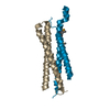
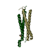
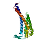
| Entry | Database: PDB / ID: 1p9y | ||||||
|---|---|---|---|---|---|---|---|
| Title | Ribosome binding of E. coli Trigger Factor mutant F44L. | ||||||
 Components Components | Trigger factor | ||||||
 Keywords Keywords | ISOMERASE / ALPHA-BETA PROTEIN | ||||||
| Function / homology |  Function and homology information Function and homology information'de novo' cotranslational protein folding / stress response to copper ion / : / protein unfolding / protein folding chaperone / peptidylprolyl isomerase / peptidyl-prolyl cis-trans isomerase activity / protein transport / ribosome binding / response to heat ...'de novo' cotranslational protein folding / stress response to copper ion / : / protein unfolding / protein folding chaperone / peptidylprolyl isomerase / peptidyl-prolyl cis-trans isomerase activity / protein transport / ribosome binding / response to heat / cell division / identical protein binding / membrane / cytosol Similarity search - Function | ||||||
| Biological species |  | ||||||
| Method |  X-RAY DIFFRACTION / SYNCHROTRON / MOLECULAR REPLACEMENT / Resolution: 2.15 Å X-RAY DIFFRACTION / SYNCHROTRON / MOLECULAR REPLACEMENT / Resolution: 2.15 Å | ||||||
 Authors Authors | Kristensen, O. / Gajhede, M. | ||||||
 Citation Citation | Journal: Structure / Year: 2003 Title: Chaperone binding at the ribosomal exit tunnel. Authors: Kristensen, O. / Gajhede, M. #1: Journal: Nature / Year: 2002Title: L23 protein functions as a chaperone docking site on the ribosome. Authors: Kramer, G. / Rauch, T. / Rist, W. / Vorderwulbecke, S. / Patzelt, H. / Schulze-Specking, A. / Ban, N. / Deuerling, E. / Bukau, B. #2: Journal: J.Mol.Biol. / Year: 2003Title: Localization of the Trigger Factor Binding Site on the Ribosomal 50S Subunit. Authors: Blaha, G. / Wilson, D.N. / Stoller, G. / Fischer, G. / Willumeit, R. / Nierhaus, K.H. #3: Journal: J.Mol.Biol. / Year: 2003Title: Interaction of Trigger Factor with the Ribosome. Authors: Maier, R. / Eckert, B. / Scholz, C. / Lilie, H. / Schmid, F.X. | ||||||
| History |
|
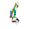
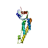
- Structure visualization
| Structure viewer | Molecule: MolmilJmol/JSmol |
|---|
- Downloads & links
-Download
| PDBx/mmCIF format | 1p9y.cif.gz | 62.5 KB | Display | PDBx/mmCIF format |
|---|---|---|---|---|
| PDB format | pdb1p9y.ent.gz | 46.4 KB | Display | PDB format |
| PDBx/mmJSON format | 1p9y.json.gz | Tree view | PDBx/mmJSON format | |
| Others |  Other downloads Other downloads |
-Validation report
| Arichive directory | https://data.pdbj.org/pub/pdb/validation_reports/p9/1p9yftp://data.pdbj.org/pub/pdb/validation_reports/p9/1p9y | HTTPS FTP |
|---|
-Related structure data
| Related structure data |  1omsSC S: Starting model for refinement C: citing same article ( |
|---|---|
| Similar structure data |


-Links
 PDBj
PDBj


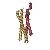
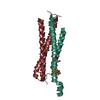
- Assembly
| Deposited unit | 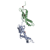
| ||||||||||||||||||
|---|---|---|---|---|---|---|---|---|---|---|---|---|---|---|---|---|---|---|---|
| 1 | 
| ||||||||||||||||||
| 2 | 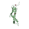
| ||||||||||||||||||
| Unit cell |
| ||||||||||||||||||
| Components on special symmetry positions |
| ||||||||||||||||||
| Details | Each of the separate chains A and B represent a biological relevant unit. |
-Components
| #1: Protein | Mass: 13330.205 Da / Num. of mol.: 2 / Fragment: Ribosome binding domain / Mutation: F44L Source method: isolated from a genetically manipulated source Source: (gene. exp.) #2: Chemical |   Mass: 60.052 Da / Num. of mol.: 3 / Source method: obtained synthetically / Formula: C2H4O2 Mass: 60.052 Da / Num. of mol.: 3 / Source method: obtained synthetically / Formula: C2H4O2#3: Water | ChemComp-HOH / |  Mass: 18.015 Da / Num. of mol.: 209 / Source method: isolated from a natural source / Formula: H2O Mass: 18.015 Da / Num. of mol.: 209 / Source method: isolated from a natural source / Formula: H2O |
|---|
-Experimental details
-Experiment
| Experiment | Method: X-RAY DIFFRACTION / Number of used crystals: 1 |
|---|
- Sample preparation
| Crystal | Density Matthews: 2.25 Å3/Da / Density % sol: 44.95 % | ||||||||||||||||||||||||||||||||||||||||||
|---|---|---|---|---|---|---|---|---|---|---|---|---|---|---|---|---|---|---|---|---|---|---|---|---|---|---|---|---|---|---|---|---|---|---|---|---|---|---|---|---|---|---|---|
| Crystal grow | Temperature: 281 K / Method: vapor diffusion, hanging drop / pH: 4.6 Details: PEG 2000 MME, AMMONIUM SULFATE, SODIUM ACETATE, TRIS, pH 4.6, VAPOR DIFFUSION, HANGING DROP, temperature 281K | ||||||||||||||||||||||||||||||||||||||||||
| Crystal grow | *PLUS Temperature: 6 ℃ / pH: 5 / Method: vapor diffusion, hanging drop | ||||||||||||||||||||||||||||||||||||||||||
| Components of the solutions | *PLUS
|
-Data collection
| Diffraction | Mean temperature: 100 K |
|---|---|
| Diffraction source | Source: SYNCHROTRON / Site: MAX II  / Beamline: I711 / Wavelength: 1.12 Å / Beamline: I711 / Wavelength: 1.12 Å |
| Detector | Type: MARRESEARCH / Detector: CCD / Date: Sep 13, 2001 |
| Radiation | Monochromator: Bendable asymmetrically cut Si(111) crystal in combination with vertically focusing cylindrical mirror. Protocol: SINGLE WAVELENGTH / Monochromatic (M) / Laue (L): M / Scattering type: x-ray |
| Radiation wavelength | Wavelength: 1.12 Å / Relative weight: 1 |
| Reflection | Resolution: 2.15→40 Å / Num. all: 14381 / Num. obs: 14381 / % possible obs: 99.7 % / Observed criterion σ(I): -3 / Redundancy: 11.3 % / Biso Wilson estimate: 17.8 Å2 / Rmerge(I) obs: 0.078 / Rsym value: 0.078 / Net I/σ(I): 25.2 |
| Reflection shell | Resolution: 2.15→2.23 Å / Redundancy: 5.9 % / Rmerge(I) obs: 0.201 / Mean I/σ(I) obs: 6.5 / Num. unique all: 1366 / Rsym value: 0.201 / % possible all: 96.7 |
| Reflection shell | *PLUS % possible obs: 96.7 % |
- Processing
| Software |
| ||||||||||||||||||||||||||||||||||||||||||||||||||||||||||||||||||||||||||||||||
|---|---|---|---|---|---|---|---|---|---|---|---|---|---|---|---|---|---|---|---|---|---|---|---|---|---|---|---|---|---|---|---|---|---|---|---|---|---|---|---|---|---|---|---|---|---|---|---|---|---|---|---|---|---|---|---|---|---|---|---|---|---|---|---|---|---|---|---|---|---|---|---|---|---|---|---|---|---|---|---|---|---|
| Refinement | Method to determine structure: MOLECULAR REPLACEMENT Starting model: PDB ENTRY 1OMS Resolution: 2.15→35.95 Å / Rfactor Rfree error: 0.007 / Data cutoff high absF: 348074.98 / Data cutoff low absF: 0 / Isotropic thermal model: RESTRAINED / Cross valid method: THROUGHOUT / σ(F): 0 / Stereochemistry target values: Engh & Huber Details: Refinement was vased on the "MLF"target function and strong NCS restraints were used THROUGHOUT. Used all data in the ultimate brief refinement cycle against a standard crystallographic target: R-FACTOR(ALL).
| ||||||||||||||||||||||||||||||||||||||||||||||||||||||||||||||||||||||||||||||||
| Solvent computation | Solvent model: FLAT MODEL / Bsol: 51.2016 Å2 / ksol: 0.382659 e/Å3 | ||||||||||||||||||||||||||||||||||||||||||||||||||||||||||||||||||||||||||||||||
| Displacement parameters | Biso mean: 27.1 Å2
| ||||||||||||||||||||||||||||||||||||||||||||||||||||||||||||||||||||||||||||||||
| Refine analyze |
| ||||||||||||||||||||||||||||||||||||||||||||||||||||||||||||||||||||||||||||||||
| Refinement step | Cycle: LAST / Resolution: 2.15→35.95 Å
| ||||||||||||||||||||||||||||||||||||||||||||||||||||||||||||||||||||||||||||||||
| Refine LS restraints |
| ||||||||||||||||||||||||||||||||||||||||||||||||||||||||||||||||||||||||||||||||
| LS refinement shell | Resolution: 2.15→2.28 Å / Rfactor Rfree error: 0.018 / Total num. of bins used: 6
| ||||||||||||||||||||||||||||||||||||||||||||||||||||||||||||||||||||||||||||||||
| Xplor file |
| ||||||||||||||||||||||||||||||||||||||||||||||||||||||||||||||||||||||||||||||||
| Refinement | *PLUS Lowest resolution: 40 Å / % reflection Rfree: 10 % / Rfactor Rwork: 0.2 | ||||||||||||||||||||||||||||||||||||||||||||||||||||||||||||||||||||||||||||||||
| Solvent computation | *PLUS | ||||||||||||||||||||||||||||||||||||||||||||||||||||||||||||||||||||||||||||||||
| Displacement parameters | *PLUS | ||||||||||||||||||||||||||||||||||||||||||||||||||||||||||||||||||||||||||||||||
| Refine LS restraints | *PLUS
|
