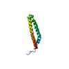
 Movie
Movie Controller


+ Open data

- Basic information
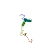
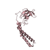
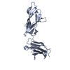
| Entry | Database: PDB / ID: 2xjz | ||||||
|---|---|---|---|---|---|---|---|
| Title | Crystal structure of the LMO2:LDB1-LID complex, C2 crystal form | ||||||
 Components Components |
| ||||||
 Keywords Keywords | ONCOPROTEIN / T-CELL LEUKEMIA / PROTO-ONCOGENE / TRANSCRIPTION / DEVELOPMENTAL PROTEIN | ||||||
| Function / homology |  Function and homology information Function and homology informationExpression and translocation of olfactory receptors / regulation of kinase activity / negative regulation of erythrocyte differentiation / cerebellar Purkinje cell differentiation / positive regulation of hemoglobin biosynthetic process / bHLH transcription factor binding / beta-catenin-TCF complex / transcription-dependent tethering of RNA polymerase II gene DNA at nuclear periphery / epithelial structure maintenance / LIM domain binding ...Expression and translocation of olfactory receptors / regulation of kinase activity / negative regulation of erythrocyte differentiation / cerebellar Purkinje cell differentiation / positive regulation of hemoglobin biosynthetic process / bHLH transcription factor binding / beta-catenin-TCF complex / transcription-dependent tethering of RNA polymerase II gene DNA at nuclear periphery / epithelial structure maintenance / LIM domain binding / gastrulation with mouth forming second / Cardiogenesis / anterior/posterior axis specification / regulation of focal adhesion assembly / cell leading edge / somatic stem cell population maintenance / hair follicle development / regulation of cell migration / positive regulation of cell adhesion / transcription coregulator binding / transcription coregulator activity / positive regulation of transcription elongation by RNA polymerase II / Wnt signaling pathway / neuron differentiation / Regulation of expression of SLITs and ROBOs / nervous system development / RUNX1 regulates transcription of genes involved in differentiation of HSCs / transcription regulator complex / transcription by RNA polymerase II / DNA-binding transcription factor binding / RNA polymerase II-specific DNA-binding transcription factor binding / transcription coactivator activity / cell adhesion / negative regulation of DNA-templated transcription / chromatin binding / chromatin / enzyme binding / negative regulation of transcription by RNA polymerase II / protein homodimerization activity / positive regulation of transcription by RNA polymerase II / protein-containing complex / DNA binding / nucleoplasm / metal ion binding / identical protein binding / nucleus Similarity search - Function | ||||||
| Biological species |  HOMO SAPIENS (human) HOMO SAPIENS (human) | ||||||
| Method |  X-RAY DIFFRACTION / SYNCHROTRON / MAD / Resolution: 2.8 Å X-RAY DIFFRACTION / SYNCHROTRON / MAD / Resolution: 2.8 Å | ||||||
 Authors Authors | El Omari, K. / Karia, D. / Porcher, C. / Mancini, E.J. | ||||||
 Citation Citation | Journal: Blood / Year: 2011 Title: Structure of the Leukemia Oncogene Lmo2: Implications for the Assembly of a Hematopoietic Transcription Factor Complex. Authors: El Omari, K. / Hoosdally, S.J. / Tuladhar, K. / Karia, D. / Vyas, P. / Patient, R. / Porcher, C. / Mancini, E.J. | ||||||
| History |
|
- Structure visualization
| Structure viewer | Molecule: MolmilJmol/JSmol |
|---|
- Downloads & links
-Download
| PDBx/mmCIF format | 2xjz.cif.gz | 327.4 KB | Display | PDBx/mmCIF format |
|---|---|---|---|---|
| PDB format | pdb2xjz.ent.gz | 270.7 KB | Display | PDB format |
| PDBx/mmJSON format | 2xjz.json.gz | Tree view | PDBx/mmJSON format | |
| Others |  Other downloads Other downloads |
-Validation report
| Arichive directory | https://data.pdbj.org/pub/pdb/validation_reports/xj/2xjzftp://data.pdbj.org/pub/pdb/validation_reports/xj/2xjz | HTTPS FTP |
|---|
-Related structure data


-Links
 PDBj
PDBj- Assembly
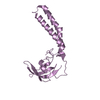
| Deposited unit | 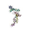
| ||||||||
|---|---|---|---|---|---|---|---|---|---|
| 1 | 
| ||||||||
| 2 | 
| ||||||||
| 3 | 
| ||||||||
| 4 | 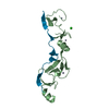
| ||||||||
| 5 | 
| ||||||||
| Unit cell |
|
-Components
| #1: Protein | Mass: 15388.909 Da / Num. of mol.: 5 / Fragment: RESIDUES 26-156 Source method: isolated from a genetically manipulated source Source: (gene. exp.) HOMO SAPIENS (human) / Production host:  #2: Protein/peptide | Mass: 3970.351 Da / Num. of mol.: 5 / Fragment: RESIDUES 334-368 Source method: isolated from a genetically manipulated source Source: (gene. exp.) HOMO SAPIENS (human) / Production host: #3: Chemical | ChemComp-ZN /   Mass: 65.409 Da / Num. of mol.: 20 / Source method: obtained synthetically / Formula: Zn Mass: 65.409 Da / Num. of mol.: 20 / Source method: obtained synthetically / Formula: Zn#4: Chemical |   Mass: 35.453 Da / Num. of mol.: 2 / Source method: obtained synthetically / Formula: Cl Mass: 35.453 Da / Num. of mol.: 2 / Source method: obtained synthetically / Formula: Cl#5: Water | ChemComp-HOH / |  Mass: 18.015 Da / Num. of mol.: 57 / Source method: isolated from a natural source / Formula: H2O Mass: 18.015 Da / Num. of mol.: 57 / Source method: isolated from a natural source / Formula: H2O |
|---|
-Experimental details
-Experiment
| Experiment | Method: X-RAY DIFFRACTION / Number of used crystals: 1 |
|---|
- Sample preparation
| Crystal | Density Matthews: 2.4 Å3/Da / Density % sol: 49 % / Description: NONE |
|---|---|
| Crystal grow | pH: 5 / Details: 1.6 M NACL, 100 MM MES PH5 AND 1 MM DTT |
-Data collection
| Diffraction | Mean temperature: 77 K |
|---|---|
| Diffraction source | Source: SYNCHROTRON / Site: ESRF  / Beamline: BM14 / Wavelength: 1.28226 / Beamline: BM14 / Wavelength: 1.28226 |
| Detector | Type: MARRESEARCH / Detector: IMAGE PLATE / Date: May 15, 2009 |
| Radiation | Protocol: MAD / Monochromatic (M) / Laue (L): M / Scattering type: x-ray |
| Radiation wavelength | Wavelength: 1.28226 Å / Relative weight: 1 |
| Reflection | Resolution: 2.8→50 Å / Num. obs: 27216 / % possible obs: 96.7 % / Observed criterion σ(I): 1.8 / Redundancy: 7.3 % / Biso Wilson estimate: 80.34 Å2 / Rmerge(I) obs: 0.14 / Net I/σ(I): 18.8 |
| Reflection shell | Resolution: 2.8→2.85 Å / Redundancy: 6.3 % / Rmerge(I) obs: 0.84 / Mean I/σ(I) obs: 1.8 / % possible all: 73.2 |
- Processing
| Software |
| |||||||||||||||||||||||||||||||||||||||||||||||||||||||||||||||||||||||||||||||||||||||||||||||||||||||||||||||||||||||||||||||||||||||||||||||||||||||||||||||||||||||||||||||||||||||||||||||||||||||||||||||||||||||||||||||||||||||||||||||||||||||||||||||||||||||||||||||||||
|---|---|---|---|---|---|---|---|---|---|---|---|---|---|---|---|---|---|---|---|---|---|---|---|---|---|---|---|---|---|---|---|---|---|---|---|---|---|---|---|---|---|---|---|---|---|---|---|---|---|---|---|---|---|---|---|---|---|---|---|---|---|---|---|---|---|---|---|---|---|---|---|---|---|---|---|---|---|---|---|---|---|---|---|---|---|---|---|---|---|---|---|---|---|---|---|---|---|---|---|---|---|---|---|---|---|---|---|---|---|---|---|---|---|---|---|---|---|---|---|---|---|---|---|---|---|---|---|---|---|---|---|---|---|---|---|---|---|---|---|---|---|---|---|---|---|---|---|---|---|---|---|---|---|---|---|---|---|---|---|---|---|---|---|---|---|---|---|---|---|---|---|---|---|---|---|---|---|---|---|---|---|---|---|---|---|---|---|---|---|---|---|---|---|---|---|---|---|---|---|---|---|---|---|---|---|---|---|---|---|---|---|---|---|---|---|---|---|---|---|---|---|---|---|---|---|---|---|---|---|---|---|---|---|---|---|---|---|---|---|---|---|---|---|---|---|---|---|---|---|---|---|---|---|---|---|---|---|---|---|---|---|---|---|---|---|---|---|---|---|---|---|---|---|---|---|---|
| Refinement | Method to determine structure: MAD Starting model: NONE Resolution: 2.8→48.32 Å / Cor.coef. Fo:Fc: 0.8949 / Cor.coef. Fo:Fc free: 0.8872 / Cross valid method: THROUGHOUT / σ(F): 0 Details: IDEAL-DIST CONTACT TERM CONTACT SETUP. RESIDUE TYPES WITHOUT CCP4 ATOM TYPE IN LIBRARY=ZN CL. NUMBER OF ATOMS WITH PROPER CCP4 ATOM TYPE=6144. NUMBER WITH APPROX DEFAULT CCP4 ATOM TYPE=0. ...Details: IDEAL-DIST CONTACT TERM CONTACT SETUP. RESIDUE TYPES WITHOUT CCP4 ATOM TYPE IN LIBRARY=ZN CL. NUMBER OF ATOMS WITH PROPER CCP4 ATOM TYPE=6144. NUMBER WITH APPROX DEFAULT CCP4 ATOM TYPE=0. NUMBER TREATED BY BAD NON-BONDED CONTACTS=22.
| |||||||||||||||||||||||||||||||||||||||||||||||||||||||||||||||||||||||||||||||||||||||||||||||||||||||||||||||||||||||||||||||||||||||||||||||||||||||||||||||||||||||||||||||||||||||||||||||||||||||||||||||||||||||||||||||||||||||||||||||||||||||||||||||||||||||||||||||||||
| Displacement parameters | Biso mean: 87.85 Å2
| |||||||||||||||||||||||||||||||||||||||||||||||||||||||||||||||||||||||||||||||||||||||||||||||||||||||||||||||||||||||||||||||||||||||||||||||||||||||||||||||||||||||||||||||||||||||||||||||||||||||||||||||||||||||||||||||||||||||||||||||||||||||||||||||||||||||||||||||||||
| Refine analyze | Luzzati coordinate error obs: 0.518 Å | |||||||||||||||||||||||||||||||||||||||||||||||||||||||||||||||||||||||||||||||||||||||||||||||||||||||||||||||||||||||||||||||||||||||||||||||||||||||||||||||||||||||||||||||||||||||||||||||||||||||||||||||||||||||||||||||||||||||||||||||||||||||||||||||||||||||||||||||||||
| Refinement step | Cycle: LAST / Resolution: 2.8→48.32 Å
| |||||||||||||||||||||||||||||||||||||||||||||||||||||||||||||||||||||||||||||||||||||||||||||||||||||||||||||||||||||||||||||||||||||||||||||||||||||||||||||||||||||||||||||||||||||||||||||||||||||||||||||||||||||||||||||||||||||||||||||||||||||||||||||||||||||||||||||||||||
| Refine LS restraints |
| |||||||||||||||||||||||||||||||||||||||||||||||||||||||||||||||||||||||||||||||||||||||||||||||||||||||||||||||||||||||||||||||||||||||||||||||||||||||||||||||||||||||||||||||||||||||||||||||||||||||||||||||||||||||||||||||||||||||||||||||||||||||||||||||||||||||||||||||||||
| LS refinement shell | Resolution: 2.8→2.91 Å / Total num. of bins used: 14
| |||||||||||||||||||||||||||||||||||||||||||||||||||||||||||||||||||||||||||||||||||||||||||||||||||||||||||||||||||||||||||||||||||||||||||||||||||||||||||||||||||||||||||||||||||||||||||||||||||||||||||||||||||||||||||||||||||||||||||||||||||||||||||||||||||||||||||||||||||
| Refinement TLS params. | Method: refined / Refine-ID: X-RAY DIFFRACTION
| |||||||||||||||||||||||||||||||||||||||||||||||||||||||||||||||||||||||||||||||||||||||||||||||||||||||||||||||||||||||||||||||||||||||||||||||||||||||||||||||||||||||||||||||||||||||||||||||||||||||||||||||||||||||||||||||||||||||||||||||||||||||||||||||||||||||||||||||||||
| Refinement TLS group |
|
