The second part of the biological assembly is generated by the crystallographic symmetry operation: -x, y+0.5, -Z+0.5
-
Components
-
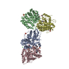
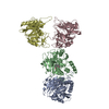
Protein , 2 types, 2 molecules AC
#1: Protein
PlateletglycoproteinIbalphachain
Mass: 31241.686 Da / Num. of mol.: 1 / Fragment: Glycoprotein 1B alpha / Mutation: N21D Source method: isolated from a genetically manipulated source Source: (gene. exp.) Homo sapiens (human) / Gene: GP1BA / Organ (production host): Ovary cells / Production host: Cricetulus griseus (Chinese hamster) / References: UniProt: P07359
#3: Protein
Prothrombin / Coagulation factor II
Mass: 29780.219 Da / Num. of mol.: 1 / Fragment: Alpha Thrombin, heavy chain / Source method: isolated from a natural source / Details: isolated from human plasma / Source: (natural) Homo sapiens (human) / References: UniProt: P00734, thrombin
-
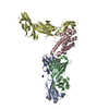
Protein/peptide / Sugars , 2 types, 2 molecules B
#2: Protein/peptide
Prothrombin / Coagulation factor II
Mass: 3432.829 Da / Num. of mol.: 1 / Fragment: Alpha Thrombin, light chain / Source method: isolated from a natural source / Details: isolated from human plasma / Source: (natural) Homo sapiens (human) / References: UniProt: P00734, thrombin
In the structure databanks used in Yorodumi, some data are registered as the other names, "COVID-19 virus" and "2019-nCoV". Here are the details of the virus and the list of structure data.
Jan 31, 2019. EMDB accession codes are about to change! (news from PDBe EMDB page)
EMDB accession codes are about to change! (news from PDBe EMDB page)
The allocation of 4 digits for EMDB accession codes will soon come to an end. Whilst these codes will remain in use, new EMDB accession codes will include an additional digit and will expand incrementally as the available range of codes is exhausted. The current 4-digit format prefixed with “EMD-” (i.e. EMD-XXXX) will advance to a 5-digit format (i.e. EMD-XXXXX), and so on. It is currently estimated that the 4-digit codes will be depleted around Spring 2019, at which point the 5-digit format will come into force.
The EM Navigator/Yorodumi systems omit the EMD- prefix.
Related info.:Q: What is EMD? / ID/Accession-code notation in Yorodumi/EM Navigator
Yorodumi is a browser for structure data from EMDB, PDB, SASBDB, etc.
This page is also the successor to EM Navigator detail page, and also detail information page/front-end page for Omokage search.
The word "yorodu" (or yorozu) is an old Japanese word meaning "ten thousand". "mi" (miru) is to see.
Related info.:EMDB / PDB / SASBDB / Comparison of 3 databanks / Yorodumi Search / Aug 31, 2016. New EM Navigator & Yorodumi / Yorodumi Papers / Jmol/JSmol / Function and homology information / Changes in new EM Navigator and Yorodumi
 Movie
Movie Controller

 Yorodumi
 Open data
 Basic information
 Components
 Keywords
 Function and homology information
 Homo sapiens (human)
 X-RAY DIFFRACTION /
  Authors
 Citation
 Structure visualization
 Downloads & links
 Other downloads


 PDBj
PDBj


 Assembly
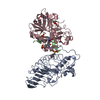


 Cricetulus griseus (Chinese hamster) / References: UniProt: P07359
 Type: D-saccharide, beta linking / Mass: 221.208 Da / Num. of mol.: 1

 Mass: 195.237 Da / Num. of mol.: 1 / Source method: obtained synthetically / Formula: C6H13NO4S / Comment: pH buffer*YM
 Mass: 166.155 Da / Num. of mol.: 1 / Source method: obtained synthetically / Formula: C6H15O3P
 Sample preparation
 / Beamline: 5.0.1 / Wavelength: 1
  Processing