 Movie
Movie Controller


+ Open data

- Basic information
| Entry | Database: PDB / ID: 1owr | ||||||
|---|---|---|---|---|---|---|---|
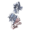
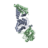
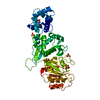
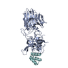
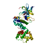
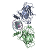
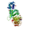
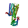
| Title | CRYSTAL STRUCTURE OF HUMAN NFAT1 BOUND MONOMERICALLY TO DNA | ||||||
 Components Components |
| ||||||
 Keywords Keywords | TRANSCRIPTION/DNA / BETA BARREL / PROTEIN-DNA COMPLEX / DOUBLE HELIX / BINARY / MONOMER / PSEUDO-CONTINUOUS / TRANSCRIPTION-DNA COMPLEX | ||||||
| Function / homology |  Function and homology information Function and homology informationtranscription factor AP-1 complex / negative regulation of vascular associated smooth muscle cell differentiation / myotube cell development / RUNX1 and FOXP3 control the development of regulatory T lymphocytes (Tregs) / calcineurin-NFAT signaling cascade / cartilage development / CLEC7A (Dectin-1) induces NFAT activation / positive regulation of myoblast fusion / Calcineurin activates NFAT / phosphatase binding ...transcription factor AP-1 complex / negative regulation of vascular associated smooth muscle cell differentiation / myotube cell development / RUNX1 and FOXP3 control the development of regulatory T lymphocytes (Tregs) / calcineurin-NFAT signaling cascade / cartilage development / CLEC7A (Dectin-1) induces NFAT activation / positive regulation of myoblast fusion / Calcineurin activates NFAT / phosphatase binding / positive regulation of B cell proliferation / 14-3-3 protein binding / cellular response to calcium ion / FCERI mediated Ca+2 mobilization / B cell receptor signaling pathway / DNA-binding transcription repressor activity, RNA polymerase II-specific / sequence-specific double-stranded DNA binding / cell migration / DNA-binding transcription activator activity, RNA polymerase II-specific / transcription regulator complex / transcription by RNA polymerase II / molecular adaptor activity / DNA-binding transcription factor activity, RNA polymerase II-specific / ribonucleoprotein complex / response to xenobiotic stimulus / RNA polymerase II cis-regulatory region sequence-specific DNA binding / DNA-binding transcription factor activity / chromatin binding / DNA damage response / regulation of transcription by RNA polymerase II / positive regulation of gene expression / positive regulation of DNA-templated transcription / chromatin / regulation of DNA-templated transcription / DNA binding / nucleoplasm / nucleus / cytoplasm / cytosol Similarity search - Function | ||||||
| Biological species |  Homo sapiens (human) Homo sapiens (human) | ||||||
| Method |  X-RAY DIFFRACTION / SYNCHROTRON / MOLECULAR REPLACEMENT / Resolution: 3 Å X-RAY DIFFRACTION / SYNCHROTRON / MOLECULAR REPLACEMENT / Resolution: 3 Å | ||||||
 Authors Authors | Stroud, J.C. / Chen, L. | ||||||
 Citation Citation | Journal: J.Mol.Biol. / Year: 2003 Title: Structure of NFAT Bound to DNA as a Monomer Authors: Stroud, J.C. / Chen, L. | ||||||
| History |
|
- Structure visualization
| Structure viewer | Molecule: MolmilJmol/JSmol |
|---|
- Downloads & links
-Download
| PDBx/mmCIF format | 1owr.cif.gz | 294.2 KB | Display | PDBx/mmCIF format |
|---|---|---|---|---|
| PDB format | pdb1owr.ent.gz | 233.8 KB | Display | PDB format |
| PDBx/mmJSON format | 1owr.json.gz | Tree view | PDBx/mmJSON format | |
| Others |  Other downloads Other downloads |
-Validation report
| Summary document | 1owr_validation.pdf.gz | 493 KB | Display | wwPDB validaton report |
|---|---|---|---|---|
| Full document | 1owr_full_validation.pdf.gz | 562.4 KB | Display | |
| Data in XML | 1owr_validation.xml.gz | 51.3 KB | Display | |
| Data in CIF | 1owr_validation.cif.gz | 70 KB | Display | |
| Arichive directory | https://data.pdbj.org/pub/pdb/validation_reports/ow/1owrftp://data.pdbj.org/pub/pdb/validation_reports/ow/1owr | HTTPS FTP |
-Related structure data
| Related structure data |  1a02S S: Starting model for refinement |
|---|---|
| Similar structure data |

-Links
 PDBj
PDBj


- Assembly
| Deposited unit | 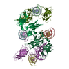
| ||||||||
|---|---|---|---|---|---|---|---|---|---|
| 1 | 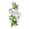
| ||||||||
| 2 | 
| ||||||||
| 3 | 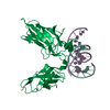
| ||||||||
| 4 | 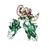
| ||||||||
| Unit cell |
| ||||||||
| Details | The biological unit is a single protein polymer of the NFAT1 RHR in complex with a double stranded piece of DNA of 13 base pairs with a 2 base overhang at the 5' end. There are 4 such biological units in the asymmetric unit. |
-Components
| #1: DNA chain | Mass: 4657.059 Da / Num. of mol.: 4 / Source method: obtained synthetically Details: The plus strand of the NFAT1 monomeric binding site was chemically synthesized #2: DNA chain | Mass: 4518.959 Da / Num. of mol.: 4 / Source method: obtained synthetically Details: The minus strand of the NFAT1 monomeric binding site was chemically synthesized #3: Protein | Mass: 32312.820 Da / Num. of mol.: 4 Source method: isolated from a genetically manipulated source Source: (gene. exp.) Homo sapiens (human) / Gene: NFATC2 OR NFAT1 OR NFATP / Production host:  |
|---|
-Experimental details
-Experiment
| Experiment | Method: X-RAY DIFFRACTION / Number of used crystals: 1 |
|---|
- Sample preparation
| Crystal | Density Matthews: 3.26 Å3/Da / Density % sol: 61.95 % | |||||||||||||||||||||||||||||||||||||||||||||||||||||||||||||||
|---|---|---|---|---|---|---|---|---|---|---|---|---|---|---|---|---|---|---|---|---|---|---|---|---|---|---|---|---|---|---|---|---|---|---|---|---|---|---|---|---|---|---|---|---|---|---|---|---|---|---|---|---|---|---|---|---|---|---|---|---|---|---|---|---|
| Crystal grow | Temperature: 289 K / Method: vapor diffusion, hanging drop / pH: 6.3 Details: sodium cacodylate, magnesium chloride, peg 3000, pH 6.3, VAPOR DIFFUSION, HANGING DROP, temperature 289K | |||||||||||||||||||||||||||||||||||||||||||||||||||||||||||||||
| Components of the solutions |
| |||||||||||||||||||||||||||||||||||||||||||||||||||||||||||||||
| Crystal grow | *PLUS Temperature: 16 ℃ / pH: 7.5 / Method: vapor diffusion, hanging drop | |||||||||||||||||||||||||||||||||||||||||||||||||||||||||||||||
| Components of the solutions | *PLUS
|
-Data collection
| Diffraction | Mean temperature: 297 K |
|---|---|
| Diffraction source | Source: SYNCHROTRON / Site: APS  / Beamline: 14-BM-C / Wavelength: 0.9 Å / Beamline: 14-BM-C / Wavelength: 0.9 Å |
| Radiation | Protocol: SINGLE WAVELENGTH / Monochromatic (M) / Laue (L): M / Scattering type: x-ray |
| Radiation wavelength | Wavelength: 0.9 Å / Relative weight: 1 |
| Reflection | Resolution: 3→20 Å / Num. all: 39696 / Num. obs: 39696 / % possible obs: 96.2 % / Observed criterion σ(F): 0 / Observed criterion σ(I): 0 / Rmerge(I) obs: 0.091 / Net I/σ(I): 15.6 |
| Reflection shell | Resolution: 3→3.07 Å / % possible all: 97.3 |
| Reflection | *PLUS Highest resolution: 3 Å / Lowest resolution: 30 Å |
| Reflection shell | *PLUS % possible obs: 97.3 % / Mean I/σ(I) obs: 2.13 |
- Processing
| Software |
| ||||||||||||||||||||
|---|---|---|---|---|---|---|---|---|---|---|---|---|---|---|---|---|---|---|---|---|---|
| Refinement | Method to determine structure: MOLECULAR REPLACEMENT Starting model: PDB ENTRY 1A02 Resolution: 3→20 Å / Isotropic thermal model: isotropic / Cross valid method: THROUGHOUT / σ(F): 0 / Stereochemistry target values: Engh & Huber Details: Starting model is the structure of the DNA binding domains of nfat, fos and jun bound to DNA without RHR-N bound to b-form dsDNA
| ||||||||||||||||||||
| Solvent computation | Bsol: 16.8187 Å2 / ksol: 0.273655 e/Å3 | ||||||||||||||||||||
| Displacement parameters | Biso mean: 66.8 Å2
| ||||||||||||||||||||
| Refine analyze |
| ||||||||||||||||||||
| Refinement step | Cycle: LAST / Resolution: 3→20 Å
| ||||||||||||||||||||
| Refine LS restraints |
| ||||||||||||||||||||
| LS refinement shell | Resolution: 3→3.19 Å / Rfactor Rfree error: 0.023
| ||||||||||||||||||||
| Refinement | *PLUS Highest resolution: 3 Å / Lowest resolution: 20 Å / % reflection Rfree: 7.7 % / Rfactor Rfree: 0.273 / Rfactor Rwork: 0.242 | ||||||||||||||||||||
| Solvent computation | *PLUS | ||||||||||||||||||||
| Displacement parameters | *PLUS | ||||||||||||||||||||
| Refine LS restraints | *PLUS
| ||||||||||||||||||||
| LS refinement shell | *PLUS Rfactor Rfree: 0.354 / Rfactor Rwork: 0.334 |
