Mass: 18.015 Da / Num. of mol.: 17 / Source method: isolated from a natural source / Formula: H2O
-
Details
Nonpolymer details
UNKNOWN ATOM OR ION (UNX): UNIDENTIFIED FEATURE ASSIGNED AS UNX UNKNOWN ATOM OR ION. UNKNOWN LIGAND ...UNKNOWN ATOM OR ION (UNX): UNIDENTIFIED FEATURE ASSIGNED AS UNX UNKNOWN ATOM OR ION. UNKNOWN LIGAND (HSE): UNIDENTIFIED LIGAND MODELED AS HOMOSERINE.
-
Experimental details
-
Experiment
Experiment
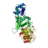
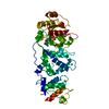
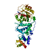
Method: X-RAY DIFFRACTION / Number of used crystals: 1
-
Sample preparation
Crystal
Density Matthews: 3.19 Å3/Da / Density % sol: 61.48 % Description: MOLECULAR REPLACEMENT MODEL WAS A MIRO STRUCTURE DETERMINED BY SAD PHASING OF SEMET-LABELED PROTEIN.
Crystal grow
pH: 8 Details: 5.0 MG/ML MIROS,1.9 M LISO4, 0.1 M BIS-TRIS PH 8.0, 5 MM EGTA; CRYSTALS WERE SOAKED IN 30 MM MGCL2, 20 MM GDP FOR 72H PRIOR TO DATA COLLECTION.
In the structure databanks used in Yorodumi, some data are registered as the other names, "COVID-19 virus" and "2019-nCoV". Here are the details of the virus and the list of structure data.
Jan 31, 2019. EMDB accession codes are about to change! (news from PDBe EMDB page)
EMDB accession codes are about to change! (news from PDBe EMDB page)
The allocation of 4 digits for EMDB accession codes will soon come to an end. Whilst these codes will remain in use, new EMDB accession codes will include an additional digit and will expand incrementally as the available range of codes is exhausted. The current 4-digit format prefixed with “EMD-” (i.e. EMD-XXXX) will advance to a 5-digit format (i.e. EMD-XXXXX), and so on. It is currently estimated that the 4-digit codes will be depleted around Spring 2019, at which point the 5-digit format will come into force.
The EM Navigator/Yorodumi systems omit the EMD- prefix.
Related info.:Q: What is EMD? / ID/Accession-code notation in Yorodumi/EM Navigator
Yorodumi is a browser for structure data from EMDB, PDB, SASBDB, etc.
This page is also the successor to EM Navigator detail page, and also detail information page/front-end page for Omokage search.
The word "yorodu" (or yorozu) is an old Japanese word meaning "ten thousand". "mi" (miru) is to see.
Related info.:EMDB / PDB / SASBDB / Comparison of 3 databanks / Yorodumi Search / Aug 31, 2016. New EM Navigator & Yorodumi / Yorodumi Papers / Jmol/JSmol / Function and homology information / Changes in new EM Navigator and Yorodumi
 Movie
Movie Controller

 Yorodumi
 Open data
 Basic information
 Components
 Keywords
 Function and homology information
 X-RAY DIFFRACTION /
  Authors
 Citation
 Structure visualization
 Downloads & links
 Other downloads


 PDBj
PDBj
 Assembly
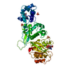


 Num. of mol.: 1 / Source method: obtained synthetically
 Mass: 96.063 Da / Num. of mol.: 3 / Source method: obtained synthetically / Formula: SO4
 Mass: 22.990 Da / Num. of mol.: 1 / Source method: obtained synthetically / Formula: Na
 Type: RNA linking / Mass: 443.201 Da / Num. of mol.: 1 / Source method: obtained synthetically / Formula: C10H15N5O11P2 / Comment: GDP, energy-carrying molecule*YM
 Mass: 24.305 Da / Num. of mol.: 2 / Source method: obtained synthetically / Formula: Mg
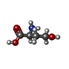
 Type: L-peptide linking / Mass: 119.119 Da / Num. of mol.: 1 / Source method: obtained synthetically / Formula: C4H9NO3
 Sample preparation
 / Beamline: 21-ID-F / Wavelength: 0.97872
  Processing