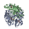
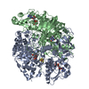
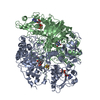
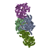
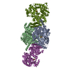
- PDB-1o95: Ternary complex between trimethylamine dehydrogenase and electron... -
+
データを開く
IDまたはキーワード:
読み込み中...
-
基本情報
登録情報
データベース: PDB / ID: 1o95
タイトル
Ternary complex between trimethylamine dehydrogenase and electron transferring flavoprotein
要素
(ELECTRON TRANSFER FLAVOPROTEIN ...) x 2
TRIMETHYLAMINE DEHYDROGENASE
キーワード
ELECTRON TRANSPORT / ELECTRON TRANSPORT-COMPLEX / PROTEIN COMPLEX / ELECTRON TRANSFER / DEHYDROGENASE / FLAVOPROTEIN / OXIDO-REDUCTASE / IRON-SULFUR / FMN
機能・相同性
機能・相同性情報
trimethylamine dehydrogenase / trimethylamine dehydrogenase activity / fatty acid beta-oxidation using acyl-CoA dehydrogenase / catabolic process / FMN binding / flavin adenine dinucleotide binding / 4 iron, 4 sulfur cluster binding / electron transfer activity / nucleotide binding / metal ion binding 類似検索 - 分子機能
Trimethylamine dehydrogenase/Dimethylamine dehydrogenase, FMN-binding domain / : / OYE-like second alpha/beta domain / : / Electron transfer flavoprotein, beta-subunit, conserved site / Electron transfer flavoprotein subunit alpha, conserved site / Electron transfer flavoprotein alpha-subunit signature. / Electron transfer flavoprotein beta-subunit signature. / Electron transfer flavoprotein, beta subunit / Electron transfer flavoprotein, alpha subunit, N-terminal ...Trimethylamine dehydrogenase/Dimethylamine dehydrogenase, FMN-binding domain / : / OYE-like second alpha/beta domain / : / Electron transfer flavoprotein, beta-subunit, conserved site / Electron transfer flavoprotein subunit alpha, conserved site / Electron transfer flavoprotein alpha-subunit signature. / Electron transfer flavoprotein beta-subunit signature. / Electron transfer flavoprotein, beta subunit / Electron transfer flavoprotein, alpha subunit, N-terminal / Electron transfer flavoprotein, beta subunit, N-terminal / Electron transfer flavoprotein domain / Electron transfer flavoprotein alpha subunit/FixB / Electron transfer flavoprotein, alpha/beta-subunit, N-terminal / Electron transfer flavoprotein, alpha subunit, C-terminal / Electron transfer flavoprotein FAD-binding domain / Electron transfer flavoprotein domain / NAD(P)-binding Rossmann-like domain / NADH:flavin oxidoreductase/NADH oxidase, N-terminal / NADH:flavin oxidoreductase / NADH oxidase family / Pyridine nucleotide-disulphide oxidoreductase / DHS-like NAD/FAD-binding domain superfamily / HUPs / FAD/NAD(P)-binding domain / FAD/NAD(P)-binding domain / Rossmann-like alpha/beta/alpha sandwich fold / 3-Layer(bba) Sandwich / FAD/NAD(P)-binding domain superfamily / Aldolase class I / Aldolase-type TIM barrel / NAD(P)-binding Rossmann-like Domain / TIM Barrel / Alpha-Beta Barrel / Rossmann fold / 3-Layer(aba) Sandwich / Alpha Beta 類似検索 - ドメイン・相同性
ADENOSINE-5'-DIPHOSPHATE / ADENOSINE MONOPHOSPHATE / FLAVIN MONONUCLEOTIDE / IRON/SULFUR CLUSTER / Trimethylamine dehydrogenase / Electron transfer flavoprotein subunit beta / Electron transfer flavoprotein subunit alpha 類似検索 - 構成要素
SHEET THE SHEET STRUCTURE OF THIS MOLECULE IS BIFURCATED. IN ORDER TO REPRESENT THIS FEATURE IN ... SHEET THE SHEET STRUCTURE OF THIS MOLECULE IS BIFURCATED. IN ORDER TO REPRESENT THIS FEATURE IN THE SHEET RECORDS BELOW, TWO SHEETS ARE DEFINED.
A: TRIMETHYLAMINE DEHYDROGENASE B: TRIMETHYLAMINE DEHYDROGENASE C: ELECTRON TRANSFER FLAVOPROTEIN BETA-SUBUNIT D: ELECTRON TRANSFER FLAVOPROTEIN ALPHA-SUBUNIT E: ELECTRON TRANSFER FLAVOPROTEIN BETA-SUBUNIT F: ELECTRON TRANSFER FLAVOPROTEIN ALPHA-SUBUNIT ヘテロ分子
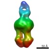
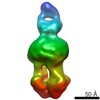
最低解像度: 3.83 Å / % possible obs: 94.3 % / Rmerge(I) obs: 0.352
-
解析
ソフトウェア
名称
バージョン
分類
REFMAC
5.1.08
精密化
DENZO
データ削減
SCALEPACK
データスケーリング
AMoRE
位相決定
精密化
構造決定の手法: 分子置換 / 解像度: 3.7→20 Å / Cor.coef. Fo:Fc: 0.884 / Cor.coef. Fo:Fc free: 0.779 / SU B: 31.707 / SU ML: 0.484 / 交差検証法: THROUGHOUT / ESU R Free: 0.991 / 立体化学のターゲット値: MAXIMUM LIKELIHOOD 詳細: DISORDERED REGIONS WERE OMITTED FROM T THE ENTIRE FAD DOMAIN FROM THE ETF MOLECULES IS NOT VISIBLE IN ELECTRON DENSITY.
Rfactor
反射数
%反射
Selection details
Rfree
0.353
1679
5 %
RANDOM
Rwork
0.252
-
-
-
obs
0.257
31940
100 %
-
溶媒の処理
イオンプローブ半径: 0.8 Å / 減衰半径: 0.8 Å / VDWプローブ半径: 1.4 Å / 溶媒モデル: BABINET MODEL WITH MASK
 ムービー
ムービー コントローラー


 データを開く
 基本情報
基本情報 要素
要素 キーワード
キーワード 機能・相同性情報
 METHYLOPHILUS METHYLOTROPHUS (バクテリア)
 X線回折 /
  データ登録者
 引用
引用 構造の表示
構造の表示 ダウンロードとリンク
 その他のダウンロード
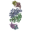


 PDBj
PDBj


 集合体
集合体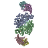


 分子量: 456.344 Da / 分子数: 2 / 由来タイプ: 合成 / 式: C17H21N4O9P
 分子量: 427.201 Da / 分子数: 2 / 由来タイプ: 合成 / 式: C10H15N5O10P2 / コメント: ADP, エネルギー貯蔵分子*YM
 分子量: 351.640 Da / 分子数: 2 / 由来タイプ: 合成 / 式: Fe4S4
 分子量: 347.221 Da / 分子数: 2 / 由来タイプ: 合成 / 式: C10H14N5O7P / コメント: AMP*YM
 試料調製
試料調製 / ビームライン: ID14-2 / 波長: 1
  解析
解析