 Movie
Movie Controller

[English] 日本語
 Yorodumi
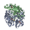
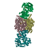
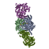
- PDB-1o94: Ternary complex between trimethylamine dehydrogenase and electron... -
+ Open data

- Basic information
| Entry | Database: PDB / ID: 1o94 | ||||||
|---|---|---|---|---|---|---|---|
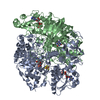
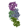
| Title | Ternary complex between trimethylamine dehydrogenase and electron transferring flavoprotein | ||||||
 Components Components |
| ||||||
 Keywords Keywords | ELECTRON TRANSPORT / PROTEIN COMPLEX / ELECTRON TRANSFER / DEHYDROGENASE | ||||||
| Function / homology |  Function and homology information Function and homology informationtrimethylamine dehydrogenase / trimethylamine dehydrogenase activity / fatty acid beta-oxidation using acyl-CoA dehydrogenase / FMN binding / flavin adenine dinucleotide binding / 4 iron, 4 sulfur cluster binding / electron transfer activity / nucleotide binding / metal ion binding Similarity search - Function | ||||||
| Biological species |  METHYLOPHILUS METHYLOTROPHUS (bacteria) METHYLOPHILUS METHYLOTROPHUS (bacteria) | ||||||
| Method |  X-RAY DIFFRACTION / SYNCHROTRON / MOLECULAR REPLACEMENT / Resolution: 2 Å X-RAY DIFFRACTION / SYNCHROTRON / MOLECULAR REPLACEMENT / Resolution: 2 Å | ||||||
 Authors Authors | Leys, D. / Basran, J. / Talfournier, F. / Sutcliffe, M.J. / Scrutton, N.S. | ||||||
 Citation Citation | Journal: Nat.Struct.Biol. / Year: 2003 Title: Extensive Conformational Sampling in a Ternary Electron Transfer Complex. Authors: Leys, D. / Basran, J. / Talfournier, F. / Sutcliffe, M.J. / Scrutton, N.S. | ||||||
| History |
| ||||||
| Remark 700 | SHEET THE SHEET STRUCTURE OF THIS MOLECULE IS BIFURCATED. IN ORDER TO REPRESENT THIS FEATURE IN ... SHEET THE SHEET STRUCTURE OF THIS MOLECULE IS BIFURCATED. IN ORDER TO REPRESENT THIS FEATURE IN THE SHEET RECORDS BELOW, TWO SHEETS ARE DEFINED. |
- Structure visualization
| Structure viewer | Molecule: MolmilJmol/JSmol |
|---|
- Downloads & links
-Download
| PDBx/mmCIF format | 1o94.cif.gz | 501.8 KB | Display | PDBx/mmCIF format |
|---|---|---|---|---|
| PDB format | pdb1o94.ent.gz | 402 KB | Display | PDB format |
| PDBx/mmJSON format | 1o94.json.gz | Tree view | PDBx/mmJSON format | |
| Others |  Other downloads Other downloads |
-Validation report
| Arichive directory | https://data.pdbj.org/pub/pdb/validation_reports/o9/1o94ftp://data.pdbj.org/pub/pdb/validation_reports/o9/1o94 | HTTPS FTP |
|---|
-Related structure data
| Related structure data | 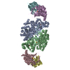 1o95C  1o96C  1o97C 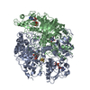 2tmdS C: citing same article ( S: Starting model for refinement |
|---|---|
| Similar structure data |


-Links
 PDBj
PDBj


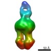
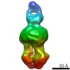
- Assembly
| Deposited unit | 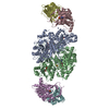
| ||||||||
|---|---|---|---|---|---|---|---|---|---|
| 1 |
| ||||||||
| Unit cell |
| ||||||||
| Components on special symmetry positions |
|
-Components
-Protein , 1 types, 2 molecules AB
| #1: Protein | Mass: 81606.023 Da / Num. of mol.: 2 / Source method: isolated from a natural source / Details: LINK BETWEEN FMN AND RESIDUE 30 / Source: (natural) METHYLOPHILUS METHYLOTROPHUS (bacteria) / References: UniProt: P16099, EC: 1.5.99.7 |
|---|
-ELECTRON TRANSFER FLAVOPROTEIN ... , 2 types, 4 molecules CEDF
| #2: Protein | Mass: 28929.850 Da / Num. of mol.: 2 Source method: isolated from a genetically manipulated source Source: (gene. exp.) METHYLOPHILUS METHYLOTROPHUS (bacteria)Production host: #3: Protein | Mass: 33622.945 Da / Num. of mol.: 2 Source method: isolated from a genetically manipulated source Source: (gene. exp.) METHYLOPHILUS METHYLOTROPHUS (bacteria)Production host: |
|---|
-Non-polymers , 5 types, 2157 molecules 


| #4: Chemical |  Mass: 456.344 Da / Num. of mol.: 2 / Source method: obtained synthetically / Formula: C17H21N4O9P Mass: 456.344 Da / Num. of mol.: 2 / Source method: obtained synthetically / Formula: C17H21N4O9P#5: Chemical |  Mass: 427.201 Da / Num. of mol.: 2 / Source method: obtained synthetically / Formula: C10H15N5O10P2 / Comment: ADP, energy-carrying molecule*YM Mass: 427.201 Da / Num. of mol.: 2 / Source method: obtained synthetically / Formula: C10H15N5O10P2 / Comment: ADP, energy-carrying molecule*YM#6: Chemical |  Mass: 351.640 Da / Num. of mol.: 2 / Source method: obtained synthetically / Formula: Fe4S4 Mass: 351.640 Da / Num. of mol.: 2 / Source method: obtained synthetically / Formula: Fe4S4#7: Chemical |  Mass: 347.221 Da / Num. of mol.: 2 / Source method: obtained synthetically / Formula: C10H14N5O7P / Comment: AMP*YM Mass: 347.221 Da / Num. of mol.: 2 / Source method: obtained synthetically / Formula: C10H14N5O7P / Comment: AMP*YM#8: Water | ChemComp-HOH / | Mass: 18.015 Da / Num. of mol.: 2149 / Source method: isolated from a natural source / Formula: H2O |
|---|
-Details
| Has protein modification | Y |
|---|
-Experimental details
-Experiment
| Experiment | Method: X-RAY DIFFRACTION / Number of used crystals: 1 |
|---|
- Sample preparation
| Crystal | Density Matthews: 2.71 Å3/Da / Density % sol: 54.55 % | ||||||||||||||||||||||||
|---|---|---|---|---|---|---|---|---|---|---|---|---|---|---|---|---|---|---|---|---|---|---|---|---|---|
| Crystal grow | pH: 6.5 Details: 8% PEG 20000, 8% PEG 750 MME, 0.1-0.3 M SODIUM ACETATE, pH 6.50 | ||||||||||||||||||||||||
| Crystal grow | *PLUS Method: vapor diffusion, hanging drop | ||||||||||||||||||||||||
| Components of the solutions | *PLUS
|
-Data collection
| Diffraction | Mean temperature: 100 K |
|---|---|
| Diffraction source | Source: SYNCHROTRON / Site: SRS  / Beamline: PX9.6 / Wavelength: 1 / Beamline: PX9.6 / Wavelength: 1 |
| Detector | Detector: CCD / Date: Apr 15, 2002 |
| Radiation | Protocol: SINGLE WAVELENGTH / Monochromatic (M) / Laue (L): M / Scattering type: x-ray |
| Radiation wavelength | Wavelength: 1 Å / Relative weight: 1 |
| Reflection | Resolution: 2→20 Å / Num. obs: 184949 / % possible obs: 94.8 % / Redundancy: 3.2 % / Rmerge(I) obs: 0.071 / Net I/σ(I): 11.5 |
| Reflection shell | Resolution: 2→2.05 Å / Rmerge(I) obs: 0.251 / Mean I/σ(I) obs: 2.9 / % possible all: 89.5 |
| Reflection | *PLUS Lowest resolution: 20 Å / Num. obs: 184250 / Num. measured all: 1099561 |
| Reflection shell | *PLUS Highest resolution: 2 Å / Lowest resolution: 2.07 Å / % possible obs: 89.5 % |
- Processing
| Software |
| ||||||||||||||||||||||||||||||||||||||||||||||||||||||||||||||||||||||||||||||||||||||||||||||||||||||||||||||||||||||||||||||||||||||||||||||||||||||||||||||||||||||||||||||||||||||
|---|---|---|---|---|---|---|---|---|---|---|---|---|---|---|---|---|---|---|---|---|---|---|---|---|---|---|---|---|---|---|---|---|---|---|---|---|---|---|---|---|---|---|---|---|---|---|---|---|---|---|---|---|---|---|---|---|---|---|---|---|---|---|---|---|---|---|---|---|---|---|---|---|---|---|---|---|---|---|---|---|---|---|---|---|---|---|---|---|---|---|---|---|---|---|---|---|---|---|---|---|---|---|---|---|---|---|---|---|---|---|---|---|---|---|---|---|---|---|---|---|---|---|---|---|---|---|---|---|---|---|---|---|---|---|---|---|---|---|---|---|---|---|---|---|---|---|---|---|---|---|---|---|---|---|---|---|---|---|---|---|---|---|---|---|---|---|---|---|---|---|---|---|---|---|---|---|---|---|---|---|---|---|---|
| Refinement | Method to determine structure: MOLECULAR REPLACEMENT Starting model: PDB ENTRY 2TMD Resolution: 2→19.88 Å / Cor.coef. Fo:Fc: 0.963 / Cor.coef. Fo:Fc free: 0.946 / SU B: 3.369 / SU ML: 0.094 / Cross valid method: THROUGHOUT / ESU R: 0.152 / ESU R Free: 0.141 / Stereochemistry target values: MAXIMUM LIKELIHOOD Details: DISORDERED REGIONS ARE NOT INCLUDED IN THE COORDINATES. THE ENTIRE FAD DOMAIN OF THE ETF MOLECULES IS NOT VISIBLE IN THE ELECTRON DENSITY.
| ||||||||||||||||||||||||||||||||||||||||||||||||||||||||||||||||||||||||||||||||||||||||||||||||||||||||||||||||||||||||||||||||||||||||||||||||||||||||||||||||||||||||||||||||||||||
| Solvent computation | Ion probe radii: 0.8 Å / Shrinkage radii: 0.8 Å / VDW probe radii: 1.4 Å / Solvent model: BABINET MODEL WITH MASK | ||||||||||||||||||||||||||||||||||||||||||||||||||||||||||||||||||||||||||||||||||||||||||||||||||||||||||||||||||||||||||||||||||||||||||||||||||||||||||||||||||||||||||||||||||||||
| Displacement parameters | Biso mean: 33.33 Å2
| ||||||||||||||||||||||||||||||||||||||||||||||||||||||||||||||||||||||||||||||||||||||||||||||||||||||||||||||||||||||||||||||||||||||||||||||||||||||||||||||||||||||||||||||||||||||
| Refinement step | Cycle: LAST / Resolution: 2→19.88 Å
| ||||||||||||||||||||||||||||||||||||||||||||||||||||||||||||||||||||||||||||||||||||||||||||||||||||||||||||||||||||||||||||||||||||||||||||||||||||||||||||||||||||||||||||||||||||||
| Refine LS restraints |
|