 Movie
Movie Controller

[English] 日本語
 Yorodumi
- PDB-1djq: STRUCTURAL AND BIOCHEMICAL CHARACTERIZATION OF RECOMBINANT C30A M... -
+ Open data

- Basic information
| Entry | Database: PDB / ID: 1djq | ||||||
|---|---|---|---|---|---|---|---|
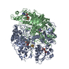
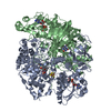
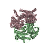
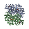
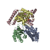
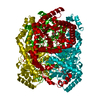
| Title | STRUCTURAL AND BIOCHEMICAL CHARACTERIZATION OF RECOMBINANT C30A MUTANT OF TRIMETHYLAMINE DEHYDROGENASE FROM METHYLOPHILUS METHYLOTROPHUS (SP. W3A1) | ||||||
 Components Components | TRIMETHYLAMINE DEHYDROGENASE | ||||||
 Keywords Keywords | OXIDOREDUCTASE / IRON-SULFUR FLAVOPROTEIN / ELECTRON TRANSFER | ||||||
| Function / homology |  Function and homology information Function and homology informationtrimethylamine dehydrogenase / trimethylamine dehydrogenase activity / FMN binding / 4 iron, 4 sulfur cluster binding / metal ion binding Similarity search - Function | ||||||
| Biological species |  Methylophilus methylotrophus (bacteria) Methylophilus methylotrophus (bacteria) | ||||||
| Method |  X-RAY DIFFRACTION / Resolution: 2.2 Å X-RAY DIFFRACTION / Resolution: 2.2 Å | ||||||
 Authors Authors | Trickey, P. / Basran, J. / Lian, L.-Y. / Chen, Z.-W. / Barton, J.D. / Sutcliffe, M.J. / Scrutton, N.S. / Mathews, F.S. | ||||||
 Citation Citation | Journal: Biochemistry / Year: 2000 Title: Structural and biochemical characterization of recombinant wild type and a C30A mutant of trimethylamine dehydrogenase from methylophilus methylotrophus (sp. W(3)A(1)). Authors: Trickey, P. / Basran, J. / Lian, L.Y. / Chen, Z. / Barton, J.D. / Sutcliffe, M.J. / Scrutton, N.S. / Mathews, F.S. #1: Journal: J.Biol.Chem. / Year: 1992Title: Correlation of X-Ray Deduced and Experimental Amino Acid Sequences of Trimethylamine Dehydrogenase Authors: Barber, M.J. / Neame, P.J. / Lim, L.W. / White, S. / Mathews, F.S. | ||||||
| History |
|
- Structure visualization
| Structure viewer | Molecule: MolmilJmol/JSmol |
|---|
- Downloads & links
-Download
| PDBx/mmCIF format | 1djq.cif.gz | 309.4 KB | Display | PDBx/mmCIF format |
|---|---|---|---|---|
| PDB format | pdb1djq.ent.gz | 247.8 KB | Display | PDB format |
| PDBx/mmJSON format | 1djq.json.gz | Tree view | PDBx/mmJSON format | |
| Others |  Other downloads Other downloads |
-Validation report
| Arichive directory | https://data.pdbj.org/pub/pdb/validation_reports/dj/1djqftp://data.pdbj.org/pub/pdb/validation_reports/dj/1djq | HTTPS FTP |
|---|
-Related structure data


-Links
 PDBj
PDBj


- Assembly
| Deposited unit | 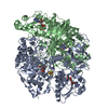
| ||||||||
|---|---|---|---|---|---|---|---|---|---|
| 1 |
| ||||||||
| Unit cell |
| ||||||||
| Details | The biological assembly is a dimer related by a molecular 2-fold axis. |
-Components
| #1: Protein | Mass: 81573.961 Da / Num. of mol.: 2 / Mutation: C30A Source method: isolated from a genetically manipulated source Source: (gene. exp.) Methylophilus methylotrophus (bacteria)Strain: W3A1 / Plasmid: PKK223-3 / Production host: #2: Chemical |   Mass: 351.640 Da / Num. of mol.: 2 / Source method: obtained synthetically / Formula: Fe4S4 Mass: 351.640 Da / Num. of mol.: 2 / Source method: obtained synthetically / Formula: Fe4S4#3: Chemical |   Mass: 456.344 Da / Num. of mol.: 2 / Source method: obtained synthetically / Formula: C17H21N4O9P Mass: 456.344 Da / Num. of mol.: 2 / Source method: obtained synthetically / Formula: C17H21N4O9P#4: Chemical |   Mass: 427.201 Da / Num. of mol.: 2 / Source method: obtained synthetically / Formula: C10H15N5O10P2 / Comment: ADP, energy-carrying molecule*YM Mass: 427.201 Da / Num. of mol.: 2 / Source method: obtained synthetically / Formula: C10H15N5O10P2 / Comment: ADP, energy-carrying molecule*YM#5: Water | ChemComp-HOH / |  Mass: 18.015 Da / Num. of mol.: 661 / Source method: isolated from a natural source / Formula: H2O Mass: 18.015 Da / Num. of mol.: 661 / Source method: isolated from a natural source / Formula: H2O |
|---|
-Experimental details
-Experiment
| Experiment | Method: X-RAY DIFFRACTION / Number of used crystals: 1 |
|---|
- Sample preparation
| Crystal | Density Matthews: 2.71 Å3/Da / Density % sol: 54.62 % |
|---|---|
| Crystal grow | Temperature: 295 K / Method: vapor diffusion, hanging drop, microseeding / pH: 6.5 Details: PEG 8000, phosphate buffer, sodium chloride, pH 6.5, vapor diffusion,hanging drop and microseeding, temperature 295.0K |
| Crystal grow | *PLUS Method: vapor diffusion, hanging drop |
-Data collection
| Diffraction | Mean temperature: 298 K |
|---|---|
| Diffraction source | Source: ROTATING ANODE / Type: RIGAKU RU200 / Wavelength: 1.5418 |
| Detector | Type: XUONG-HAMLIN MULTIWIRE / Detector: AREA DETECTOR / Date: Nov 21, 1995 |
| Radiation | Protocol: SINGLE WAVELENGTH / Monochromatic (M) / Laue (L): M / Scattering type: x-ray |
| Radiation wavelength | Wavelength: 1.5418 Å / Relative weight: 1 |
| Reflection | Resolution: 2.22→10 Å / Num. all: 86826 / Num. obs: 81611 / % possible obs: 93.4 % / Observed criterion σ(F): 0 / Observed criterion σ(I): 0 / Redundancy: 4 % / Biso Wilson estimate: 18.6 Å2 / Rmerge(I) obs: 0.055 / Net I/σ(I): 22.2 |
| Reflection shell | Resolution: 2.22→2.32 Å / Redundancy: 2.4 % / Rmerge(I) obs: 0.123 / Num. unique all: 17184 / % possible all: 71.7 |
| Reflection | *PLUS Lowest resolution: 50 Å |
| Reflection shell | *PLUS % possible obs: 71.7 % / Mean I/σ(I) obs: 6.4 |
- Processing
| Software |
| |||||||||||||||||||||||||
|---|---|---|---|---|---|---|---|---|---|---|---|---|---|---|---|---|---|---|---|---|---|---|---|---|---|---|
| Refinement | Resolution: 2.2→8 Å / σ(F): 2 / Stereochemistry target values: Engh & Huber
| |||||||||||||||||||||||||
| Refinement step | Cycle: LAST / Resolution: 2.2→8 Å
| |||||||||||||||||||||||||
| Refine LS restraints |
|
