 Movie
Movie Controller

[English] 日本語
 Yorodumi
- PDB-1nsq: MECHANISM OF PHOSPHATE TRANSFER BY NUCLEOSIDE DIPHOSPHATE KINASE:... -
+ Open data

- Basic information
| Entry | Database: PDB / ID: 1nsq | ||||||
|---|---|---|---|---|---|---|---|
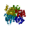
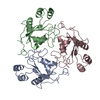
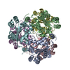
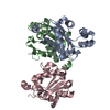
| Title | MECHANISM OF PHOSPHATE TRANSFER BY NUCLEOSIDE DIPHOSPHATE KINASE: X-RAY STRUCTURES OF A PHOSPHO-HISTIDINE INTERMEDIATE OF THE ENZYMES FROM DROSOPHILA AND DICTYOSTELIUM | ||||||
 Components Components | NUCLEOSIDE DIPHOSPHATE KINASE Nucleoside-diphosphate kinase Nucleoside-diphosphate kinase | ||||||
 Keywords Keywords | PHOSPHOTRANSFERASE | ||||||
| Function / homology |  Function and homology information Function and homology informationregulation of tube architecture, open tracheal system / Azathioprine ADME / Ribavirin ADME / establishment or maintenance of polarity of follicular epithelium / open tracheal system development / epithelial cell migration, open tracheal system / Interconversion of nucleotide di- and triphosphates / nuclear microtubule / Neutrophil degranulation / nucleoside-diphosphate kinase ...regulation of tube architecture, open tracheal system / Azathioprine ADME / Ribavirin ADME / establishment or maintenance of polarity of follicular epithelium / open tracheal system development / epithelial cell migration, open tracheal system / Interconversion of nucleotide di- and triphosphates / nuclear microtubule / Neutrophil degranulation / nucleoside-diphosphate kinase / nucleotide metabolic process / UTP biosynthetic process / CTP biosynthetic process / GTP biosynthetic process / adherens junction organization / nucleoside diphosphate kinase activity / microtubule-based process / mitotic cell cycle / kinase activity / microtubule binding / microtubule / phosphorylation / GTP binding / magnesium ion binding / ATP binding / plasma membrane / cytoplasmSimilarity search - Function | ||||||
| Biological species |  Drosophila melanogaster (fruit fly) Drosophila melanogaster (fruit fly) | ||||||
| Method | X-RAY DIFFRACTION / Resolution: 2.18 Å | ||||||
 Authors Authors | Janin, J. / Chiadmi, M. / Morera, S. / Lebras, G. / Lascu, I. | ||||||
 Citation Citation | Journal: Biochemistry / Year: 1995 Title: Mechanism of phosphate transfer by nucleoside diphosphate kinase: X-ray structures of the phosphohistidine intermediate of the enzymes from Drosophila and Dictyostelium. Authors: Morera, S. / Chiadmi, M. / LeBras, G. / Lascu, I. / Janin, J. #1: Journal: Structure / Year: 1993Title: Crystal Structure of the Awd Nucleoside Diphosphate Kinase from Drosophila Authors: Chiadmi, M. / Morera, S. / Lascu, I. / Dumas, C. / Lebras, G. / Veron, M. / Janin, J. #2: Journal: Embo J. / Year: 1992Title: X-Ray Structure of Nucleoside Diphosphate Kinase Authors: Dumas, C. / Lascu, I. / Morera, S. / Glaser, P. / Fourme, R. / Wallet, V. / Lacombe, M.L. / Veron, M. / Janin, J. | ||||||
| History |
|
- Structure visualization
| Structure viewer | Molecule: MolmilJmol/JSmol |
|---|
- Downloads & links
-Download
| PDBx/mmCIF format | 1nsq.cif.gz | 99.1 KB | Display | PDBx/mmCIF format |
|---|---|---|---|---|
| PDB format | pdb1nsq.ent.gz | 82.8 KB | Display | PDB format |
| PDBx/mmJSON format | 1nsq.json.gz | Tree view | PDBx/mmJSON format | |
| Others |  Other downloads Other downloads |
-Validation report
| Arichive directory | https://data.pdbj.org/pub/pdb/validation_reports/ns/1nsqftp://data.pdbj.org/pub/pdb/validation_reports/ns/1nsq | HTTPS FTP |
|---|
-Related structure data


-Links
 PDBj
PDBj

- Assembly
| Deposited unit | 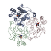
| ||||||||
|---|---|---|---|---|---|---|---|---|---|
| 1 | 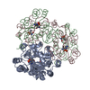
| ||||||||
| Unit cell |
|
-Components
| #1: Protein | Nucleoside-diphosphate kinase Mass: 17270.830 Da / Num. of mol.: 3 Source method: isolated from a genetically manipulated source Source: (gene. exp.) Drosophila melanogaster (fruit fly) / References: UniProt: P08879, nucleoside-diphosphate kinase#2: Water | ChemComp-HOH / | Water Mass: 18.015 Da / Num. of mol.: 257 / Source method: isolated from a natural source / Formula: H2O Mass: 18.015 Da / Num. of mol.: 257 / Source method: isolated from a natural source / Formula: H2O |
|---|
-Experimental details
-Experiment
| Experiment | Method: X-RAY DIFFRACTION |
|---|
- Sample preparation
| Crystal | Density Matthews: 3.68 Å3/Da / Density % sol: 66.53 % | ||||||||||||||||||||||||||||||
|---|---|---|---|---|---|---|---|---|---|---|---|---|---|---|---|---|---|---|---|---|---|---|---|---|---|---|---|---|---|---|---|
| Crystal grow | *PLUS Temperature: 18 ℃ / pH: 8 / Method: vapor diffusion, hanging drop | ||||||||||||||||||||||||||||||
| Components of the solutions | *PLUS
|
-Data collection
| Radiation | Scattering type: x-ray |
|---|---|
| Radiation wavelength | Relative weight: 1 |
| Reflection | Resolution: 2.17→15.1 Å / Num. obs: 32198 / % possible obs: 79 % |
| Reflection | *PLUS Num. measured all: 184822 / Rmerge(I) obs: 0.077 |
- Processing
| Software |
| ||||||||||||||||||||||||||||||||||||||||||||||||||||||||||||
|---|---|---|---|---|---|---|---|---|---|---|---|---|---|---|---|---|---|---|---|---|---|---|---|---|---|---|---|---|---|---|---|---|---|---|---|---|---|---|---|---|---|---|---|---|---|---|---|---|---|---|---|---|---|---|---|---|---|---|---|---|---|
| Refinement | Resolution: 2.18→8 Å / σ(F): 2 /
| ||||||||||||||||||||||||||||||||||||||||||||||||||||||||||||
| Refinement step | Cycle: LAST / Resolution: 2.18→8 Å
| ||||||||||||||||||||||||||||||||||||||||||||||||||||||||||||
| Refine LS restraints |
| ||||||||||||||||||||||||||||||||||||||||||||||||||||||||||||
| Software | *PLUS Name: X-PLOR / Classification: refinement | ||||||||||||||||||||||||||||||||||||||||||||||||||||||||||||
| Refinement | *PLUS Num. reflection obs: 31522 | ||||||||||||||||||||||||||||||||||||||||||||||||||||||||||||
| Solvent computation | *PLUS | ||||||||||||||||||||||||||||||||||||||||||||||||||||||||||||
| Displacement parameters | *PLUS Biso mean: 22 Å2 |
