- PDB-1ndl: THE AWD NUCLEOTIDE DIPHOSPHATE KINASE FROM DROSOPHILA -
+
Open data
ID or keywords:
Loading...
-
Basic information
Entry
Database: PDB / ID: 1ndl
Title
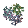
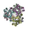
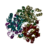
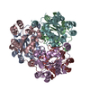
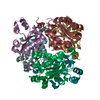
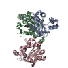
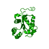
THE AWD NUCLEOTIDE DIPHOSPHATE KINASE FROM DROSOPHILA
Components
NUCLEOSIDE DIPHOSPHATE KINASE
Keywords
PHOSPHOTRANSFERASE
Function / homology
Function and homology information
ribonucleoside triphosphate biosynthetic process / regulation of tube architecture, open tracheal system / Azathioprine ADME / Ribavirin ADME / establishment or maintenance of polarity of follicular epithelium / epithelial cell migration, open tracheal system / Interconversion of nucleotide di- and triphosphates / deoxyribonucleoside triphosphate biosynthetic process / open tracheal system development / Neutrophil degranulation ...ribonucleoside triphosphate biosynthetic process / regulation of tube architecture, open tracheal system / Azathioprine ADME / Ribavirin ADME / establishment or maintenance of polarity of follicular epithelium / epithelial cell migration, open tracheal system / Interconversion of nucleotide di- and triphosphates / deoxyribonucleoside triphosphate biosynthetic process / open tracheal system development / Neutrophil degranulation / nuclear microtubule / nucleoside-diphosphate kinase / adherens junction organization / UTP biosynthetic process / nucleotide metabolic process / CTP biosynthetic process / nucleoside diphosphate kinase activity / GTP biosynthetic process / microtubule-based process / kinase activity / mitotic cell cycle / microtubule binding / microtubule / GTP binding / magnesium ion binding / mitochondrion / ATP binding / plasma membrane Similarity search - Function
In the structure databanks used in Yorodumi, some data are registered as the other names, "COVID-19 virus" and "2019-nCoV". Here are the details of the virus and the list of structure data.
Jan 31, 2019. EMDB accession codes are about to change! (news from PDBe EMDB page)
EMDB accession codes are about to change! (news from PDBe EMDB page)
The allocation of 4 digits for EMDB accession codes will soon come to an end. Whilst these codes will remain in use, new EMDB accession codes will include an additional digit and will expand incrementally as the available range of codes is exhausted. The current 4-digit format prefixed with “EMD-” (i.e. EMD-XXXX) will advance to a 5-digit format (i.e. EMD-XXXXX), and so on. It is currently estimated that the 4-digit codes will be depleted around Spring 2019, at which point the 5-digit format will come into force.
The EM Navigator/Yorodumi systems omit the EMD- prefix.
Related info.:Q: What is EMD? / ID/Accession-code notation in Yorodumi/EM Navigator
Yorodumi is a browser for structure data from EMDB, PDB, SASBDB, etc.
This page is also the successor to EM Navigator detail page, and also detail information page/front-end page for Omokage search.
The word "yorodu" (or yorozu) is an old Japanese word meaning "ten thousand". "mi" (miru) is to see.
Related info.:EMDB / PDB / SASBDB / Comparison of 3 databanks / Yorodumi Search / Aug 31, 2016. New EM Navigator & Yorodumi / Yorodumi Papers / Jmol/JSmol / Function and homology information / Changes in new EM Navigator and Yorodumi
 Movie
Movie Controller


 Open data
 Basic information
 Components
 Keywords
 Function and homology information
 X-RAY DIFFRACTION / Resolution: 2.4 Å
  Authors
 Citation
 Structure visualization
 Downloads & links
 Other downloads
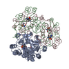

 PDBj
PDBj

 Assembly
 Mass: 18.015 Da / Num. of mol.: 315 / Source method: isolated from a natural source / Formula: H2O
 Sample preparation
 Processing