 Movie
Movie Controller


+ Open data

- Basic information
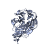
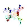
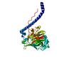
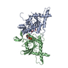
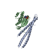
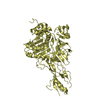
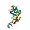
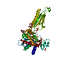
| Entry | Database: PDB / ID: 1npe | ||||||
|---|---|---|---|---|---|---|---|
| Title | Crystal structure of Nidogen/Laminin Complex | ||||||
 Components Components |
| ||||||
 Keywords Keywords | STRUCTURAL PROTEIN / Glycoprotein / Basement membrane / beta-propeller / EGF-like | ||||||
| Function / homology |  Function and homology information Function and homology informationLaminin interactions / laminin-511 trimer / tissue morphogenesis / hair follicle cell proliferation / regulation of basement membrane organization / glomerular basement membrane development / hemidesmosome assembly / laminin-1 binding / glycosphingolipid binding / positive regulation of integrin-mediated signaling pathway ...Laminin interactions / laminin-511 trimer / tissue morphogenesis / hair follicle cell proliferation / regulation of basement membrane organization / glomerular basement membrane development / hemidesmosome assembly / laminin-1 binding / glycosphingolipid binding / positive regulation of integrin-mediated signaling pathway / Degradation of the extracellular matrix / hair cell differentiation / protein complex involved in cell-matrix adhesion / extracellular matrix binding / proteoglycan binding / positive regulation of cell-substrate adhesion / extracellular matrix structural constituent / hair follicle morphogenesis / positive regulation of muscle cell differentiation / basement membrane / extracellular matrix disassembly / synaptic cleft / collagen binding / laminin binding / extracellular matrix organization / positive regulation of cell adhesion / cell-matrix adhesion / cell periphery / neuromuscular junction / extracellular matrix / : / neuron projection development / cell migration / chromatin organization / protein-containing complex assembly / gene expression / cell adhesion / calcium ion binding / extracellular region Similarity search - Function | ||||||
| Biological species |  | ||||||
| Method |  X-RAY DIFFRACTION / SYNCHROTRON / MOLECULAR REPLACEMENT / Resolution: 2.3 Å X-RAY DIFFRACTION / SYNCHROTRON / MOLECULAR REPLACEMENT / Resolution: 2.3 Å | ||||||
 Authors Authors | Takagi, J. / Yang, Y.T. / Liu, J.-H. / Wang, J.-H. / Springer, T.A. | ||||||
 Citation Citation | Journal: Nature / Year: 2003 Title: Complex between nidogen and laminin fragments reveals a paradigmatic beta-propeller interface Authors: Takagi, J. / Yang, Y.T. / Liu, J.-H. / Wang, J.-H. / Springer, T.A. | ||||||
| History |
|
- Structure visualization
| Structure viewer | Molecule: MolmilJmol/JSmol |
|---|
- Downloads & links
-Download
| PDBx/mmCIF format | 1npe.cif.gz | 99.7 KB | Display | PDBx/mmCIF format |
|---|---|---|---|---|
| PDB format | pdb1npe.ent.gz | 74.7 KB | Display | PDB format |
| PDBx/mmJSON format | 1npe.json.gz | Tree view | PDBx/mmJSON format | |
| Others |  Other downloads Other downloads |
-Validation report
| Arichive directory | https://data.pdbj.org/pub/pdb/validation_reports/np/1npeftp://data.pdbj.org/pub/pdb/validation_reports/np/1npe | HTTPS FTP |
|---|
-Related structure data
-Links
 PDBj
PDBj


- Assembly
| Deposited unit | 
| ||||||||
|---|---|---|---|---|---|---|---|---|---|
| 1 |
| ||||||||
| Unit cell |
|
-Components
| #1: Protein | Mass: 29998.078 Da / Num. of mol.: 1 Fragment: G3 YWTD domain, sequence database residue 941-1203 Source method: isolated from a genetically manipulated source Source: (gene. exp.)  Homo sapiens (human) / References: UniProt: P02468, UniProt: P10493*PLUS Homo sapiens (human) / References: UniProt: P02468, UniProt: P10493*PLUS | ||||
|---|---|---|---|---|---|
| #2: Protein | Mass: 17366.607 Da / Num. of mol.: 1 Fragment: modules III 3-5, sequence database residue 769-932 Source method: isolated from a genetically manipulated source Source: (gene. exp.) Homo sapiens (human) / References: UniProt: P10493, UniProt: P02468*PLUS | ||||
| #3: Chemical | ChemComp-CD /   Mass: 112.411 Da / Num. of mol.: 6 / Source method: obtained synthetically / Formula: Cd Mass: 112.411 Da / Num. of mol.: 6 / Source method: obtained synthetically / Formula: Cd#4: Water | ChemComp-HOH / |  Mass: 18.015 Da / Num. of mol.: 127 / Source method: isolated from a natural source / Formula: H2O Mass: 18.015 Da / Num. of mol.: 127 / Source method: isolated from a natural source / Formula: H2OHas protein modification | Y | |
-Experimental details
-Experiment
| Experiment | Method: X-RAY DIFFRACTION / Number of used crystals: 1 |
|---|
- Sample preparation
| Crystal | Density Matthews: 3 Å3/Da / Density % sol: 58.64 % | |||||||||||||||||||||||||||||||||||
|---|---|---|---|---|---|---|---|---|---|---|---|---|---|---|---|---|---|---|---|---|---|---|---|---|---|---|---|---|---|---|---|---|---|---|---|---|
| Crystal grow | Temperature: 298 K / Method: vapor diffusion, hanging drop / pH: 7.5 Details: 1 M NaAc, 0.05 M CdSO4, 0.1 M, HEPES pH 7.5, VAPOR DIFFUSION, HANGING DROP, temperature 298K | |||||||||||||||||||||||||||||||||||
| Crystal grow | *PLUS Method: vapor diffusion, hanging drop | |||||||||||||||||||||||||||||||||||
| Components of the solutions | *PLUS
|
-Data collection
| Diffraction | Mean temperature: 100 K |
|---|---|
| Diffraction source | Source: SYNCHROTRON / Site: APS  / Beamline: 19-ID / Wavelength: 0.9537 Å / Beamline: 19-ID / Wavelength: 0.9537 Å |
| Detector | Type: ADSC QUANTUM 4 / Detector: CCD / Date: Aug 1, 2002 |
| Radiation | Monochromator: graphite / Protocol: SINGLE WAVELENGTH / Monochromatic (M) / Laue (L): M / Scattering type: x-ray |
| Radiation wavelength | Wavelength: 0.9537 Å / Relative weight: 1 |
| Reflection | Resolution: 2.3→50 Å / Num. all: 25891 / Num. obs: 25885 / % possible obs: 99 % / Observed criterion σ(F): 0 / Observed criterion σ(I): 0 / Redundancy: 18 % / Biso Wilson estimate: 33.1 Å2 / Rmerge(I) obs: 0.105 / Net I/σ(I): 16.7 |
| Reflection shell | Resolution: 2.3→2.38 Å / Redundancy: 1.8 % / Rmerge(I) obs: 0.424 / Mean I/σ(I) obs: 3.4 / Num. unique all: 2375 / % possible all: 92.5 |
| Reflection | *PLUS Lowest resolution: 20 Å / Num. obs: 25940 / % possible obs: 99 % / Num. measured all: 466114 |
| Reflection shell | *PLUS % possible obs: 92.5 % / Mean I/σ(I) obs: 2.7 |
- Processing
| Software |
| |||||||||||||||||||||||||
|---|---|---|---|---|---|---|---|---|---|---|---|---|---|---|---|---|---|---|---|---|---|---|---|---|---|---|
| Refinement | Method to determine structure: MOLECULAR REPLACEMENT Starting model: pdb entry 1IJQ and 1KLO Resolution: 2.3→20 Å / Cross valid method: THROUGHOUT / σ(F): 0
| |||||||||||||||||||||||||
| Displacement parameters | Biso mean: 27.2 Å2
| |||||||||||||||||||||||||
| Refine analyze |
| |||||||||||||||||||||||||
| Refinement step | Cycle: LAST / Resolution: 2.3→20 Å
| |||||||||||||||||||||||||
| Refine LS restraints |
| |||||||||||||||||||||||||
| Refinement | *PLUS Rfactor Rfree: 0.257 / Rfactor Rwork: 0.226 | |||||||||||||||||||||||||
| Solvent computation | *PLUS | |||||||||||||||||||||||||
| Displacement parameters | *PLUS | |||||||||||||||||||||||||
| Refine LS restraints | *PLUS
|
