 Movie
Movie Controller


+ Open data

- Basic information
| Entry | Database: PDB / ID: 1k2f | ||||||
|---|---|---|---|---|---|---|---|
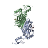
| Title | siah, Seven In Absentia Homolog | ||||||
 Components Components | siah-1A protein | ||||||
 Keywords Keywords | LIGASE / PROTEIN BINDING / beta-sandwich | ||||||
| Function / homology |  Function and homology information Function and homology informationbeta-catenin destruction complex / Antigen processing: Ubiquitination & Proteasome degradation / ubiquitin conjugating enzyme binding / SCF ubiquitin ligase complex / male meiosis I / regulation of multicellular organism growth / canonical Wnt signaling pathway / positive regulation of intrinsic apoptotic signaling pathway / protein catabolic process / post-embryonic development ...beta-catenin destruction complex / Antigen processing: Ubiquitination & Proteasome degradation / ubiquitin conjugating enzyme binding / SCF ubiquitin ligase complex / male meiosis I / regulation of multicellular organism growth / canonical Wnt signaling pathway / positive regulation of intrinsic apoptotic signaling pathway / protein catabolic process / post-embryonic development / RING-type E3 ubiquitin transferase / protein destabilization / disordered domain specific binding / ubiquitin-protein transferase activity / ubiquitin protein ligase activity / neuron apoptotic process / spermatogenesis / ubiquitin-dependent protein catabolic process / proteasome-mediated ubiquitin-dependent protein catabolic process / early endosome / cell differentiation / protein ubiquitination / positive regulation of apoptotic process / protein homodimerization activity / zinc ion binding / identical protein binding / nucleus / cytoplasm / cytosol Similarity search - Function | ||||||
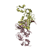
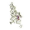
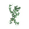
| Biological species |  | ||||||
| Method |  X-RAY DIFFRACTION / MAD / Resolution: 2.6 Å X-RAY DIFFRACTION / MAD / Resolution: 2.6 Å | ||||||
 Authors Authors | Polekhina, G. / House, C.M. / Traficante, N. / Mackay, J.P. / Relaix, F. / Sassoon, D.A. / Parker, M.W. / Bowtell, D.D.L. | ||||||
 Citation Citation | Journal: Nat.Struct.Biol. / Year: 2002 Title: Siah ubiquitin ligase is structurally related to TRAF and modulates TNF-alpha signaling. Authors: Polekhina, G. / House, C.M. / Traficante, N. / Mackay, J.P. / Relaix, F. / Sassoon, D.A. / Parker, M.W. / Bowtell, D.D. #1: Journal: DEVELOPMENT / Year: 1993Title: Isolation and characterisation of murine homologues of the Drosophila seven inabsentia gene (sina) Authors: Della, N.G. / Senior, P.V. / Bowtell, D.D. | ||||||
| History |
|
- Structure visualization
| Structure viewer | Molecule: MolmilJmol/JSmol |
|---|
- Downloads & links
-Download
| PDBx/mmCIF format | 1k2f.cif.gz | 87.4 KB | Display | PDBx/mmCIF format |
|---|---|---|---|---|
| PDB format | pdb1k2f.ent.gz | 67 KB | Display | PDB format |
| PDBx/mmJSON format | 1k2f.json.gz | Tree view | PDBx/mmJSON format | |
| Others |  Other downloads Other downloads |
-Validation report
| Arichive directory | https://data.pdbj.org/pub/pdb/validation_reports/k2/1k2fftp://data.pdbj.org/pub/pdb/validation_reports/k2/1k2f | HTTPS FTP |
|---|
-Related structure data
| Similar structure data |
|---|


-Links
 PDBj
PDBj


- Assembly
| Deposited unit | 
| ||||||||
|---|---|---|---|---|---|---|---|---|---|
| 1 |
| ||||||||
| 2 | 
| ||||||||
| Unit cell |
|
-Components
| #1: Protein | Mass: 21423.533 Da / Num. of mol.: 2 / Fragment: C-terminal domain (residues 93-282) Source method: isolated from a genetically manipulated source Source: (gene. exp.)  #2: Chemical | ChemComp-ZN /   Mass: 65.409 Da / Num. of mol.: 6 / Source method: obtained synthetically / Formula: Zn Mass: 65.409 Da / Num. of mol.: 6 / Source method: obtained synthetically / Formula: Zn#3: Chemical | ChemComp-BME /   Mass: 78.133 Da / Num. of mol.: 4 / Source method: obtained synthetically / Formula: C2H6OS Mass: 78.133 Da / Num. of mol.: 4 / Source method: obtained synthetically / Formula: C2H6OS#4: Water | ChemComp-HOH / |  Mass: 18.015 Da / Num. of mol.: 65 / Source method: isolated from a natural source / Formula: H2O Mass: 18.015 Da / Num. of mol.: 65 / Source method: isolated from a natural source / Formula: H2OHas protein modification | N | |
|---|
-Experimental details
-Experiment
| Experiment | Method: X-RAY DIFFRACTION / Number of used crystals: 1 |
|---|
- Sample preparation
| Crystal | Density Matthews: 2.31 Å3/Da / Density % sol: 46.75 % | ||||||||||||||||||||||||||||
|---|---|---|---|---|---|---|---|---|---|---|---|---|---|---|---|---|---|---|---|---|---|---|---|---|---|---|---|---|---|
| Crystal grow | Temperature: 295 K / Method: vapor diffusion, hanging drop / pH: 6.5 Details: calcium chloride, ethanol, MES, pH 6.5, VAPOR DIFFUSION, HANGING DROP, temperature 295K | ||||||||||||||||||||||||||||
| Crystal grow | *PLUS | ||||||||||||||||||||||||||||
| Components of the solutions | *PLUS
|
-Data collection
| Diffraction | Mean temperature: 100 K |
|---|---|
| Diffraction source | Source: ROTATING ANODE / Type: RIGAKU / Wavelength: 1.54 Å |
| Detector | Type: MARRESEARCH / Detector: IMAGE PLATE / Date: Nov 13, 2000 / Details: mirrors |
| Radiation | Protocol: SINGLE WAVELENGTH / Monochromatic (M) / Laue (L): M / Scattering type: x-ray |
| Radiation wavelength | Wavelength: 1.54 Å / Relative weight: 1 |
| Reflection | Resolution: 2.6→20 Å / Num. all: 12661 / Num. obs: 12661 / % possible obs: 99.8 % / Observed criterion σ(F): 0 / Observed criterion σ(I): 0 / Redundancy: 8 % / Biso Wilson estimate: 64.8 Å2 / Rmerge(I) obs: 0.063 / Rsym value: 0.063 / Net I/σ(I): 28 |
| Reflection shell | Resolution: 2.6→2.69 Å / Redundancy: 6 % / Rmerge(I) obs: 0.388 / Mean I/σ(I) obs: 4.31 / Num. unique all: 1244 / Rsym value: 0.388 / % possible all: 99.4 |
| Reflection | *PLUS |
- Processing
| Software |
| ||||||||||||||||||||||||||||||
|---|---|---|---|---|---|---|---|---|---|---|---|---|---|---|---|---|---|---|---|---|---|---|---|---|---|---|---|---|---|---|---|
| Refinement | Method to determine structure: MAD / Resolution: 2.6→20 Å / Isotropic thermal model: Isotropic / Cross valid method: THROUGHOUT / σ(F): 0 / σ(I): 0 / Stereochemistry target values: Engh & Huber
| ||||||||||||||||||||||||||||||
| Displacement parameters | Biso mean: 56.7 Å2 | ||||||||||||||||||||||||||||||
| Refinement step | Cycle: LAST / Resolution: 2.6→20 Å
| ||||||||||||||||||||||||||||||
| Software | *PLUS Name: CNS / Version: 1 / Classification: refinement | ||||||||||||||||||||||||||||||
| Refinement | *PLUS Highest resolution: 2.6 Å / Lowest resolution: 20 Å / σ(F): 0 / % reflection Rfree: 5 % / Rfactor obs: 0.228 | ||||||||||||||||||||||||||||||
| Solvent computation | *PLUS | ||||||||||||||||||||||||||||||
| Displacement parameters | *PLUS Biso mean: 56.7 Å2 | ||||||||||||||||||||||||||||||
| Refine LS restraints | *PLUS
|
