Mass: 18.015 Da / Num. of mol.: 83 / Source method: isolated from a natural source / Formula: H2O
-
Details
Has protein modification
Y
-
Experimental details
-
Experiment
Experiment
Method: X-RAY DIFFRACTION / Number of used crystals: 4
-
Sample preparation
Crystal
Density Matthews: 2.72 Å3/Da / Density % sol: 54.79 %
Crystal grow
Temperature: 300 K / Method: vapor diffusion, hanging drop / pH: 5.6 Details: ammonium sulphate, PEG 3350, sodium citrate, pH 5.6, VAPOR DIFFUSION, HANGING DROP, temperature 300K
Crystal grow
*PLUS
pH: 8
Components of the solutions
*PLUS
ID
Conc.
Common name
Crystal-ID
Sol-ID
Details
1
8mg/ml
protein
1
drop
2
5mM
Tris-HCl
1
drop
pH8.0
3
50mM
ammoniumsulfate
1
reservoir
4
18 %
PEG3350
1
reservoir
5
2.5mM
sodiumcitrate
1
reservoir
pH5.6
-
Data collection
Diffraction
ID
Mean temperature (K)
Crystal-ID
1
100
1
2
100
1
3
100
1
4
100
1
Diffraction source
Source
Site
Beamline
Type
ID
Wavelength
Wavelength (Å)
ROTATING ANODE
MACSCIENCE
1
1.5418
1.5418
ROTATING ANODE
RIGAKU RUH3R
2
1.5418
1.5418
SYNCHROTRON
APS
14-BM-C
3
1
1
Detector
Type
ID
Detector
Details
RIGAKU RAXIS II
1
IMAGE PLATE
YaleMirrors
RIGAKU RAXIS IV
2
IMAGE PLATE
Monocapillaryoptics
ADSC QUANTUM 4
3
CCD
APSBM14Coptics
Radiation
ID
Monochromator
Protocol
Monochromatic (M) / Laue (L)
Scattering type
Wavelength-ID
1
Yalemirrors
SINGLEWAVELENGTH
M
x-ray
1
2
Monocapillaryoptics
SINGLEWAVELENGTH
M
x-ray
1
3
Monocapillaryoptics
SINGLEWAVELENGTH
M
x-ray
1
4
APSBM14C
SINGLEWAVELENGTH
M
x-ray
1
Radiation wavelength
ID
Wavelength (Å)
Relative weight
1
1.5418
1
2
1
1
Reflection
Resolution: 2.4→50 Å / % possible obs: 99.4 % / Observed criterion σ(I): 1 / Redundancy: 18.6 % / Biso Wilson estimate: 58.3 Å2 / Rmerge(I) obs: 0.11 / Rsym value: 0.07 / Net I/σ(I): 39
Reflection shell
Resolution: 2.35→2.4 Å / Rmerge(I) obs: 0.57 / Mean I/σ(I) obs: 3.7 / % possible all: 100
Reflection
*PLUS
Lowest resolution: 50 Å
Reflection shell
*PLUS
% possible obs: 100 %
-
Processing
Software
Name
Version
Classification
DENZO
datareduction
SCALEPACK
datascaling
SHARP
phasing
CNS
1
refinement
Refinement
Method to determine structure: MIR Starting model: ab initio built into MIR map Resolution: 2.4→6 Å / Cross valid method: THROUGHOUT / σ(F): 0 / σ(I): 0 / Stereochemistry target values: Engh & Huber
Rfactor
Num. reflection
% reflection
Selection details
Rfree
0.29
731
-
Random
Rwork
0.22
-
-
-
all
0.22
15630
-
-
obs
0.22
15298
97.9 %
-
Displacement parameters
Baniso -1
Baniso -2
Baniso -3
1-
-6.369 Å2
0 Å2
0 Å2
2-
-
-6.369 Å2
0 Å2
3-
-
-
12.737 Å2
Refinement step
Cycle: LAST / Resolution: 2.4→6 Å
Protein
Nucleic acid
Ligand
Solvent
Total
Num. atoms
2360
0
137
83
2580
Refine LS restraints
Refine-ID
Type
Dev ideal
Dev ideal target
X-RAY DIFFRACTION
c_bond_d
0.015
X-RAY DIFFRACTION
c_angle_deg
1.96
X-RAY DIFFRACTION
c_mcbond_it
6.394
3.5
X-RAY DIFFRACTION
c_mcangle_it
7.619
4
X-RAY DIFFRACTION
c_scbond_it
7.922
4
X-RAY DIFFRACTION
c_scangle_it
9.105
4.5
Xplor file
Refine-ID
Serial no
Param file
Topol file
X-RAY DIFFRACTION
1
protein_rep.param
protein.top
X-RAY DIFFRACTION
2
carbohydrate.param
carbohydrate.top
X-RAY DIFFRACTION
3
water_rep.param
water.top
X-RAY DIFFRACTION
4
ion.param
ion.top
Refinement
*PLUS
Solvent computation
*PLUS
Displacement parameters
*PLUS
+
About Yorodumi
-
News
-
Feb 9, 2022. New format data for meta-information of EMDB entries
New format data for meta-information of EMDB entries
Version 3 of the EMDB header file is now the official format.
The previous official version 1.9 will be removed from the archive.
In the structure databanks used in Yorodumi, some data are registered as the other names, "COVID-19 virus" and "2019-nCoV". Here are the details of the virus and the list of structure data.
Jan 31, 2019. EMDB accession codes are about to change! (news from PDBe EMDB page)
EMDB accession codes are about to change! (news from PDBe EMDB page)
The allocation of 4 digits for EMDB accession codes will soon come to an end. Whilst these codes will remain in use, new EMDB accession codes will include an additional digit and will expand incrementally as the available range of codes is exhausted. The current 4-digit format prefixed with “EMD-” (i.e. EMD-XXXX) will advance to a 5-digit format (i.e. EMD-XXXXX), and so on. It is currently estimated that the 4-digit codes will be depleted around Spring 2019, at which point the 5-digit format will come into force.
The EM Navigator/Yorodumi systems omit the EMD- prefix.
Related info.:Q: What is EMD? / ID/Accession-code notation in Yorodumi/EM Navigator
Yorodumi is a browser for structure data from EMDB, PDB, SASBDB, etc.
This page is also the successor to EM Navigator detail page, and also detail information page/front-end page for Omokage search.
The word "yorodu" (or yorozu) is an old Japanese word meaning "ten thousand". "mi" (miru) is to see.
Related info.:EMDB / PDB / SASBDB / Comparison of 3 databanks / Yorodumi Search / Aug 31, 2016. New EM Navigator & Yorodumi / Yorodumi Papers / Jmol/JSmol / Function and homology information / Changes in new EM Navigator and Yorodumi
 Movie
Movie Controller

 Yorodumi
 Open data
 Basic information
 Components
 Keywords
 Function and homology information
 Homo sapiens (human)
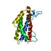
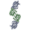
 X-RAY DIFFRACTION /
  Authors
 Citation
 Structure visualization
 Downloads & links
 Other downloads


 PDBj
PDBj


 Assembly


 Cricetulus griseus (Chinese hamster) / Strain (production host): CHO / References: UniProt: P08887
 Type: D-saccharide, beta linking / Mass: 221.208 Da / Num. of mol.: 2


 Mass: 96.063 Da / Num. of mol.: 2 / Source method: obtained synthetically / Formula: SO4
 Type: L-peptide linking / Mass: 121.158 Da / Num. of mol.: 1 / Source method: obtained synthetically / Formula: C3H7NO2S
 Sample preparation
 Processing