 Movie
Movie Controller


+ Open data

- Basic information
| Entry | Database: PDB / ID: 1i1r | ||||||
|---|---|---|---|---|---|---|---|
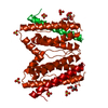
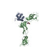
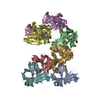
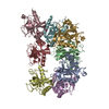
| Title | CRYSTAL STRUCTURE OF A CYTOKINE/RECEPTOR COMPLEX | ||||||
 Components Components |
| ||||||
 Keywords Keywords | CYTOKINE / cytokine-receptor complex / gp130 / viral IL-6 | ||||||
| Function / homology |  Function and homology information Function and homology informationtype I oncostatin-M receptor complex / interleukin-27 receptor activity / oncostatin-M-mediated signaling pathway / ciliary neurotrophic factor receptor activity / ciliary neurotrophic factor receptor binding / leukemia inhibitory factor signaling pathway / interleukin-11 receptor activity / interleukin-11 binding / interleukin-6 receptor activity / negative regulation of interleukin-6-mediated signaling pathway ...type I oncostatin-M receptor complex / interleukin-27 receptor activity / oncostatin-M-mediated signaling pathway / ciliary neurotrophic factor receptor activity / ciliary neurotrophic factor receptor binding / leukemia inhibitory factor signaling pathway / interleukin-11 receptor activity / interleukin-11 binding / interleukin-6 receptor activity / negative regulation of interleukin-6-mediated signaling pathway / ciliary neurotrophic factor receptor complex / ciliary neurotrophic factor-mediated signaling pathway / interleukin-6 receptor complex / interleukin-27-mediated signaling pathway / interleukin-11-mediated signaling pathway / interleukin-6 receptor binding / T-helper 17 cell lineage commitment / positive regulation of acute inflammatory response / positive regulation of astrocyte differentiation / positive regulation of adaptive immune response / intestinal epithelial cell development / positive regulation of platelet aggregation / IL-6-type cytokine receptor ligand interactions / Interleukin-27 signaling / Interleukin-35 Signalling / cell surface receptor signaling pathway via STAT / cytokine receptor activity / Interleukin-6 signaling / glycogen metabolic process / interleukin-6-mediated signaling pathway / growth factor binding / positive regulation of Notch signaling pathway / MAPK3 (ERK1) activation / positive regulation of cardiac muscle hypertrophy / MAPK1 (ERK2) activation / cytokine binding / protein tyrosine kinase activator activity / positive regulation of vascular endothelial growth factor production / positive regulation of osteoblast differentiation / coreceptor activity / response to cytokine / positive regulation of T cell proliferation / cytokine activity / cytokine-mediated signaling pathway / scaffold protein binding / negative regulation of neuron apoptotic process / signaling receptor complex / immune response / membrane raft / external side of plasma membrane / neuronal cell body / positive regulation of cell population proliferation / dendrite / negative regulation of apoptotic process / : / extracellular exosome / extracellular region / membrane / identical protein binding / plasma membrane Similarity search - Function | ||||||
| Biological species |  Homo sapiens (human) Homo sapiens (human)  Human herpesvirus 8 Human herpesvirus 8 | ||||||
| Method |  X-RAY DIFFRACTION / SYNCHROTRON / MOLECULAR REPLACEMENT / Resolution: 2.4 Å X-RAY DIFFRACTION / SYNCHROTRON / MOLECULAR REPLACEMENT / Resolution: 2.4 Å | ||||||
 Authors Authors | Chow, D. / He, X. / Snow, A.L. / Rose-John, S. / Garcia, K.C. | ||||||
 Citation Citation | Journal: Science / Year: 2001 Title: Structure of an extracellular gp130 cytokine receptor signaling complex. Authors: Chow, D. / He, X. / Snow, A.L. / Rose-John, S. / Garcia, K.C. | ||||||
| History |
| ||||||
| Remark 300 | BIOMOLECULE: 1 THIS ENTRY CONTAINS THE CRYSTALLOGRAPHIC ASYMMETRIC UNIT WHICH CONSISTS OF 2 CHAIN(S) ...BIOMOLECULE: 1 THIS ENTRY CONTAINS THE CRYSTALLOGRAPHIC ASYMMETRIC UNIT WHICH CONSISTS OF 2 CHAIN(S). SEE REMARK 350 FOR INFORMATION ON GENERATING THE BIOLOGICAL MOLECULE(S). THE BIOLOGICAL ASSEMBLY IS A TETRAMER, OF WHICH HALF (ONE VIL-6, ONE GP130) IS IN THE ASYMMETRIC UNIT. THE DYAD-AXIS OF THE TETRAMER IS THE C2 CRYSTALLOGRAPHIC AXIS. NOTE: COORDINATES FOR THE ENTIRE TETRAMER CAN BE OBTAINED DIRECTLY FROM THE AUTHORS (kcgarcia@stanford.edu). |
- Structure visualization
| Structure viewer | Molecule: MolmilJmol/JSmol |
|---|
- Downloads & links
-Download
| PDBx/mmCIF format | 1i1r.cif.gz | 115.7 KB | Display | PDBx/mmCIF format |
|---|---|---|---|---|
| PDB format | pdb1i1r.ent.gz | 88.8 KB | Display | PDB format |
| PDBx/mmJSON format | 1i1r.json.gz | Tree view | PDBx/mmJSON format | |
| Others |  Other downloads Other downloads |
-Validation report
| Arichive directory | https://data.pdbj.org/pub/pdb/validation_reports/i1/1i1rftp://data.pdbj.org/pub/pdb/validation_reports/i1/1i1r | HTTPS FTP |
|---|
-Related structure data


-Links
 PDBj
PDBj


- Assembly
| Deposited unit | 
| ||||||||
|---|---|---|---|---|---|---|---|---|---|
| 1 | 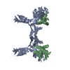
| ||||||||
| Unit cell |
| ||||||||
| Details | The second part of the biological assembly is generated by the 2-fold crystallographic axis |
-Components
| #1: Protein | Mass: 34664.105 Da / Num. of mol.: 1 Fragment: DOMAINS 1, 2, 3 OF THE GP130 EXTRACELLULAR DOMAIN (RESIDUES 1-303) Source method: isolated from a genetically manipulated source Details: EXPRESSED IN THE PRESENCE OF INHIBITOR OF N-LINKED GLYCOSYLATION (TUNICAMYCIN) Source: (gene. exp.) Homo sapiens (human) / Plasmid: PACGP67A / Cell line (production host): SF9 / Production host:   Spodoptera frugiperda (fall armyworm) / References: UniProt: P40189 Spodoptera frugiperda (fall armyworm) / References: UniProt: P40189 |
|---|---|
| #2: Protein | Mass: 20857.197 Da / Num. of mol.: 1 Source method: isolated from a genetically manipulated source Details: NON-GLYCOSYLATED / Source: (gene. exp.) Human herpesvirus 8 / Genus: Rhadinovirus / Plasmid: PACGP67A / Cell line (production host): SF9 / Production host: Spodoptera frugiperda (fall armyworm) / References: UniProt: Q98823 |
| #3: Water | ChemComp-HOH /  Mass: 18.015 Da / Num. of mol.: 370 / Source method: isolated from a natural source / Formula: H2O Mass: 18.015 Da / Num. of mol.: 370 / Source method: isolated from a natural source / Formula: H2O |
| Has protein modification | Y |
-Experimental details
-Experiment
| Experiment | Method: X-RAY DIFFRACTION / Number of used crystals: 1 |
|---|
- Sample preparation
| Crystal | Density Matthews: 4.07 Å3/Da / Density % sol: 69.77 % | ||||||||||||||||||||
|---|---|---|---|---|---|---|---|---|---|---|---|---|---|---|---|---|---|---|---|---|---|
| Crystal grow | Temperature: 293 K / Method: vapor diffusion, sitting drop / pH: 6.5 Details: MPEG 2000, sodium citrate, pH 6.5, VAPOR DIFFUSION, SITTING DROP, temperature 293K | ||||||||||||||||||||
| Crystal | *PLUS Density % sol: 70 % | ||||||||||||||||||||
| Crystal grow | *PLUS Method: unknown | ||||||||||||||||||||
| Components of the solutions | *PLUS
|
-Data collection
| Diffraction | Mean temperature: 100 K |
|---|---|
| Diffraction source | Source: SYNCHROTRON / Site: ALS  / Beamline: 5.0.2 / Wavelength: 1 Å / Beamline: 5.0.2 / Wavelength: 1 Å |
| Detector | Type: ADSC QUANTUM 4 / Detector: CCD / Date: Sep 21, 2000 |
| Radiation | Protocol: SINGLE WAVELENGTH / Monochromatic (M) / Laue (L): M / Scattering type: x-ray |
| Radiation wavelength | Wavelength: 1 Å / Relative weight: 1 |
| Reflection | Resolution: 2.4→50 Å / Num. all: 34376 / Num. obs: 34376 / % possible obs: 99 % / Observed criterion σ(F): 0 / Observed criterion σ(I): 0 / Redundancy: 3.1 % / Biso Wilson estimate: 50 Å2 / Rmerge(I) obs: 0.081 / Net I/σ(I): 3.4 |
| Reflection shell | Resolution: 2.4→2.49 Å / Redundancy: 3.1 % / Rmerge(I) obs: 0.487 / Mean I/σ(I) obs: 1.4 / Num. unique all: 3420 / % possible all: 99.6 |
| Reflection | *PLUS Num. measured all: 106397 |
| Reflection shell | *PLUS % possible obs: 99.6 % |
- Processing
| Software |
| |||||||||||||||||||||||||
|---|---|---|---|---|---|---|---|---|---|---|---|---|---|---|---|---|---|---|---|---|---|---|---|---|---|---|
| Refinement | Method to determine structure: MOLECULAR REPLACEMENT Starting model: human Interleukin-6 1ALU, gp130 D2D3 domains 1BQU Resolution: 2.4→50 Å / Isotropic thermal model: Isotropic / Cross valid method: THROUGHOUT / σ(F): 0 / σ(I): 0 / Stereochemistry target values: Engh & Huber / Details: wilson B value 50.2
| |||||||||||||||||||||||||
| Displacement parameters | Biso mean: 49.1 Å2
| |||||||||||||||||||||||||
| Refine analyze |
| |||||||||||||||||||||||||
| Refinement step | Cycle: LAST / Resolution: 2.4→50 Å
| |||||||||||||||||||||||||
| Refine LS restraints |
| |||||||||||||||||||||||||
| LS refinement shell | Resolution: 2.4→2.49 Å / Rfactor Rfree error: 0.019
|
