 Movie
Movie Controller

[English] 日本語
 Yorodumi
- PDB-5zqe: Crystal Structure of Penicillin-Binding Protein D2 from Listeria ... -
+ Open data

- Basic information
| Entry | Database: PDB / ID: 5zqe | ||||||
|---|---|---|---|---|---|---|---|
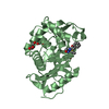
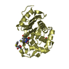
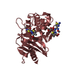
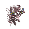
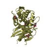
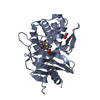
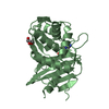
| Title | Crystal Structure of Penicillin-Binding Protein D2 from Listeria monocytogenes in the Cefuroxime bound form | ||||||
 Components Components | Lmo2812 protein | ||||||
 Keywords Keywords | ANTIBIOTIC / penicillin-binding protein D2 / Listeria monocytogenes / antibiotics / beta-lactams / DD-carboxypeptidase / lmo2812 / LmPBPD2 | ||||||
| Function / homology |  Function and homology information Function and homology informationserine-type D-Ala-D-Ala carboxypeptidase activity / peptidoglycan biosynthetic process / cell wall organization / regulation of cell shape / proteolysis / metal ion binding Similarity search - Function | ||||||
| Biological species |  Listeria monocytogenes EGD-e (bacteria) Listeria monocytogenes EGD-e (bacteria) | ||||||
| Method |  X-RAY DIFFRACTION / SYNCHROTRON / MOLECULAR REPLACEMENT / Resolution: 1.996 Å X-RAY DIFFRACTION / SYNCHROTRON / MOLECULAR REPLACEMENT / Resolution: 1.996 Å | ||||||
 Authors Authors | Jeong, J.H. / Kim, Y.G. | ||||||
| Funding support |  Korea, Republic Of, 1items Korea, Republic Of, 1items
| ||||||
 Citation Citation | Journal: Antimicrob. Agents Chemother. / Year: 2018 Title: Crystal Structures of Penicillin-Binding Protein D2 from Listeria monocytogenes and Structural Basis for Antibiotic Specificity Authors: Jeong, J.H. / Cha, H.J. / Kim, Y.G. | ||||||
| History |
|
- Structure visualization
| Structure viewer | Molecule: MolmilJmol/JSmol |
|---|
- Downloads & links
-Download
| PDBx/mmCIF format | 5zqe.cif.gz | 401.1 KB | Display | PDBx/mmCIF format |
|---|---|---|---|---|
| PDB format | pdb5zqe.ent.gz | 323.8 KB | Display | PDB format |
| PDBx/mmJSON format | 5zqe.json.gz | Tree view | PDBx/mmJSON format | |
| Others |  Other downloads Other downloads |
-Validation report
| Arichive directory | https://data.pdbj.org/pub/pdb/validation_reports/zq/5zqeftp://data.pdbj.org/pub/pdb/validation_reports/zq/5zqe | HTTPS FTP |
|---|
-Related structure data
| Related structure data | 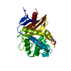 5zqaSC  5zqbC  5zqcC 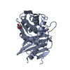 5zqdC S: Starting model for refinement C: citing same article ( |
|---|---|
| Similar structure data |


-Links
 PDBj
PDBj


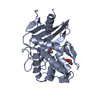
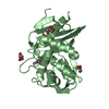
- Assembly
| Deposited unit | 
| ||||||||
|---|---|---|---|---|---|---|---|---|---|
| 1 | 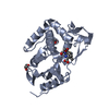
| ||||||||
| 2 | 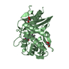
| ||||||||
| 3 | 
| ||||||||
| 4 | 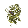
| ||||||||
| 5 | 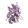
| ||||||||
| 6 | 
| ||||||||
| 7 | 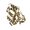
| ||||||||
| 8 | 
| ||||||||
| Unit cell |
|
-Components
| #1: Protein | Mass: 30091.605 Da / Num. of mol.: 8 / Fragment: UNP residues 21-272 Source method: isolated from a genetically manipulated source Details: it contains covalently bound cefuroxime molecules Source: (gene. exp.) Listeria monocytogenes EGD-e (bacteria)Strain: EGD-e / Gene: lmo2812 / Production host: #2: Chemical | ChemComp-CES /   Mass: 383.376 Da / Num. of mol.: 8 / Source method: obtained synthetically / Formula: C15H17N3O7S / Feature type: SUBJECT OF INVESTIGATION Mass: 383.376 Da / Num. of mol.: 8 / Source method: obtained synthetically / Formula: C15H17N3O7S / Feature type: SUBJECT OF INVESTIGATION#3: Chemical | ChemComp-GOL /   Mass: 92.094 Da / Num. of mol.: 16 / Source method: obtained synthetically / Formula: C3H8O3 Mass: 92.094 Da / Num. of mol.: 16 / Source method: obtained synthetically / Formula: C3H8O3#4: Chemical | ChemComp-PEG / |   Mass: 106.120 Da / Num. of mol.: 1 / Source method: obtained synthetically / Formula: C4H10O3 Mass: 106.120 Da / Num. of mol.: 1 / Source method: obtained synthetically / Formula: C4H10O3#5: Water | ChemComp-HOH / |  Mass: 18.015 Da / Num. of mol.: 921 / Source method: isolated from a natural source / Formula: H2O Mass: 18.015 Da / Num. of mol.: 921 / Source method: isolated from a natural source / Formula: H2OHas protein modification | Y | |
|---|
-Experimental details
-Experiment
| Experiment | Method: X-RAY DIFFRACTION / Number of used crystals: 1 |
|---|
- Sample preparation
| Crystal | Density Matthews: 2.33 Å3/Da / Density % sol: 47.17 % |
|---|---|
| Crystal grow | Temperature: 295 K / Method: vapor diffusion, sitting drop / pH: 7 |
-Data collection
| Diffraction | Mean temperature: 200 K |
|---|---|
| Diffraction source | Source: SYNCHROTRON / Site: PAL/PLS / Beamline: 5C (4A) / Wavelength: 0.9794 Å |
| Detector | Type: ADSC QUANTUM 315r / Detector: CCD / Date: Jul 23, 2016 |
| Radiation | Monochromator: Double crystal SI(111) / Protocol: SINGLE WAVELENGTH / Monochromatic (M) / Laue (L): M / Scattering type: x-ray |
| Radiation wavelength | Wavelength: 0.9794 Å / Relative weight: 1 |
| Reflection | Resolution: 1.996→29.982 Å / Num. obs: 149025 / % possible obs: 91.13 % / Observed criterion σ(I): 0 / Redundancy: 6.1 % / Biso Wilson estimate: 13.29 Å2 / Rsym value: 0.127 / Net I/σ(I): 11.7 |
| Reflection shell | Resolution: 1.996→2.067 Å / Redundancy: 5.9 % / Num. unique obs: 14478 / Rsym value: 0.475 / % possible all: 84.24 |
- Processing
| Software |
| |||||||||||||||||||||||||||||||||||||||||||||||||||||||||||||||||||||||||||||||||||||||||||||||||||||||||
|---|---|---|---|---|---|---|---|---|---|---|---|---|---|---|---|---|---|---|---|---|---|---|---|---|---|---|---|---|---|---|---|---|---|---|---|---|---|---|---|---|---|---|---|---|---|---|---|---|---|---|---|---|---|---|---|---|---|---|---|---|---|---|---|---|---|---|---|---|---|---|---|---|---|---|---|---|---|---|---|---|---|---|---|---|---|---|---|---|---|---|---|---|---|---|---|---|---|---|---|---|---|---|---|---|---|---|
| Refinement | Method to determine structure: MOLECULAR REPLACEMENT Starting model: 5ZQA Resolution: 1.996→29.982 Å / Cross valid method: NONE / σ(F): 0 / Phase error: 38.41
| |||||||||||||||||||||||||||||||||||||||||||||||||||||||||||||||||||||||||||||||||||||||||||||||||||||||||
| Solvent computation | Shrinkage radii: 0.9 Å / VDW probe radii: 1.11 Å | |||||||||||||||||||||||||||||||||||||||||||||||||||||||||||||||||||||||||||||||||||||||||||||||||||||||||
| Refinement step | Cycle: LAST / Resolution: 1.996→29.982 Å
| |||||||||||||||||||||||||||||||||||||||||||||||||||||||||||||||||||||||||||||||||||||||||||||||||||||||||
| Refine LS restraints |
| |||||||||||||||||||||||||||||||||||||||||||||||||||||||||||||||||||||||||||||||||||||||||||||||||||||||||
| LS refinement shell |
|
