 Movie
Movie Controller

[English] 日本語
 Yorodumi
- PDB-1m9d: X-ray crystal structure of Cyclophilin A/HIV-1 CA N-terminal doma... -
+ Open data

- Basic information
| Entry | Database: PDB / ID: 1m9d | ||||||
|---|---|---|---|---|---|---|---|
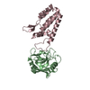
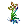
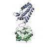
| Title | X-ray crystal structure of Cyclophilin A/HIV-1 CA N-terminal domain (1-146) O-type chimera Complex. | ||||||
 Components Components |
| ||||||
 Keywords Keywords | Isomerase/Viral protein / CAPSID / HIV-1 / CYCLOPHILIN A / ISOMERASE / ROTAMASE / Isomerase-Viral protein COMPLEX | ||||||
| Function / homology |  Function and homology information Function and homology informationnegative regulation of protein K48-linked ubiquitination / regulation of apoptotic signaling pathway / cell adhesion molecule production / lipid droplet organization / negative regulation of viral life cycle / heparan sulfate binding / regulation of viral genome replication / virion binding / leukocyte chemotaxis / negative regulation of stress-activated MAPK cascade ...negative regulation of protein K48-linked ubiquitination / regulation of apoptotic signaling pathway / cell adhesion molecule production / lipid droplet organization / negative regulation of viral life cycle / heparan sulfate binding / regulation of viral genome replication / virion binding / leukocyte chemotaxis / negative regulation of stress-activated MAPK cascade / activation of protein kinase B activity / endothelial cell activation / Basigin interactions / protein peptidyl-prolyl isomerization / cyclosporin A binding / Minus-strand DNA synthesis / Plus-strand DNA synthesis / Uncoating of the HIV Virion / viral budding via host ESCRT complex / Early Phase of HIV Life Cycle / Integration of provirus / APOBEC3G mediated resistance to HIV-1 infection / negative regulation of protein phosphorylation / viral release from host cell / Calcineurin activates NFAT / Binding and entry of HIV virion / negative regulation of oxidative stress-induced intrinsic apoptotic signaling pathway / negative regulation of protein kinase activity / positive regulation of viral genome replication / neutrophil chemotaxis / Gene and protein expression by JAK-STAT signaling after Interleukin-12 stimulation / positive regulation of protein secretion / HIV-1 retropepsin / peptidylprolyl isomerase / symbiont-mediated activation of host apoptosis / retroviral ribonuclease H / peptidyl-prolyl cis-trans isomerase activity / exoribonuclease H / exoribonuclease H activity / Assembly Of The HIV Virion / : / Budding and maturation of HIV virion / DNA integration / platelet activation / viral genome integration into host DNA / establishment of integrated proviral latency / RNA-directed DNA polymerase / platelet aggregation / integrin binding / RNA stem-loop binding / viral penetration into host nucleus / host multivesicular body / RNA-directed DNA polymerase activity / RNA-DNA hybrid ribonuclease activity / positive regulation of protein phosphorylation / neuron differentiation / Transferases; Transferring phosphorus-containing groups; Nucleotidyltransferases / SARS-CoV-1 activates/modulates innate immune responses / unfolded protein binding / Platelet degranulation / host cell / protein folding / viral nucleocapsid / cellular response to oxidative stress / secretory granule lumen / DNA recombination / vesicle / DNA-directed DNA polymerase / ficolin-1-rich granule lumen / aspartic-type endopeptidase activity / Hydrolases; Acting on ester bonds / DNA-directed DNA polymerase activity / positive regulation of MAPK cascade / symbiont-mediated suppression of host gene expression / viral translational frameshifting / focal adhesion / apoptotic process / Neutrophil degranulation / symbiont entry into host cell / lipid binding / host cell nucleus / host cell plasma membrane / virion membrane / structural molecule activity / protein-containing complex / proteolysis / extracellular space / DNA binding / RNA binding / extracellular exosome / extracellular region / zinc ion binding / membrane / nucleus / cytoplasm / cytosol Similarity search - Function | ||||||
| Biological species |  Homo sapiens (human) Homo sapiens (human)  Human immunodeficiency virus 1 Human immunodeficiency virus 1 | ||||||
| Method |  X-RAY DIFFRACTION / SYNCHROTRON / MOLECULAR REPLACEMENT / Resolution: 1.9 Å X-RAY DIFFRACTION / SYNCHROTRON / MOLECULAR REPLACEMENT / Resolution: 1.9 Å | ||||||
 Authors Authors | Howard, B.R. / Vajdos, F.F. / Li, S. / Sundquist, W.I. / Hill, C.P. | ||||||
 Citation Citation | Journal: Nat.Struct.Biol. / Year: 2003 Title: Structural insights into the catalytic mechanism of cyclophilin A Authors: Howard, B.R. / Vajdos, F.F. / Li, S. / Sundquist, W.I. / Hill, C.P. | ||||||
| History |
| ||||||
| Remark 300 | BIOMOLECULE: 1, 2 THIS ENTRY CONTAINS THE CRYSTALLOGRAPHIC ASYMMETRIC UNIT WHICH CONSISTS OF 4 ...BIOMOLECULE: 1, 2 THIS ENTRY CONTAINS THE CRYSTALLOGRAPHIC ASYMMETRIC UNIT WHICH CONSISTS OF 4 CHAIN(S). SEE REMARK 350 FOR INFORMATION ON GENERATING THE BIOLOGICAL MOLECULE(S). Complex "A" consists of chains B and C; Complex "B" consists of chains A and D. | ||||||
| Remark 999 | SEQUENCE According to the authors, this apparent conflict is due to the use of HIV-1 strain NL4-3 ...SEQUENCE According to the authors, this apparent conflict is due to the use of HIV-1 strain NL4-3 which has a histidine at residue 120. |
- Structure visualization
| Structure viewer | Molecule: MolmilJmol/JSmol |
|---|
- Downloads & links
-Download
| PDBx/mmCIF format | 1m9d.cif.gz | 141.1 KB | Display | PDBx/mmCIF format |
|---|---|---|---|---|
| PDB format | pdb1m9d.ent.gz | 111.9 KB | Display | PDB format |
| PDBx/mmJSON format | 1m9d.json.gz | Tree view | PDBx/mmJSON format | |
| Others |  Other downloads Other downloads |
-Validation report
| Arichive directory | https://data.pdbj.org/pub/pdb/validation_reports/m9/1m9dftp://data.pdbj.org/pub/pdb/validation_reports/m9/1m9d | HTTPS FTP |
|---|
-Related structure data
| Related structure data |  1m9cC  1m9eC  1m9fC  1m9xC  1m9yC 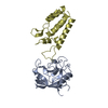 1ak4S  1m96 S: Starting model for refinement C: citing same article ( |
|---|---|
| Similar structure data |


-Links
 PDBj
PDBj


- Assembly
| Deposited unit | 
| ||||||||
|---|---|---|---|---|---|---|---|---|---|
| 1 | 
| ||||||||
| 2 | 
| ||||||||
| Unit cell |
|
-Components
| #1: Protein | Mass: 18036.504 Da / Num. of mol.: 2 Source method: isolated from a genetically manipulated source Source: (gene. exp.) Homo sapiens (human) / Plasmid: PET3A / Species (production host): Escherichia coli / Production host:  #2: Protein | Mass: 16191.552 Da / Num. of mol.: 2 / Fragment: N-TERMINAL DOMAIN / Mutation: L83T, V86P, H87A, A88M, I91L, A92P, M96I Source method: isolated from a genetically manipulated source Source: (gene. exp.) Human immunodeficiency virus 1 / Genus: Lentivirus / Gene: CA / Plasmid: PET11A / Species (production host): Escherichia coli / Production host: #3: Water | ChemComp-HOH / |  Mass: 18.015 Da / Num. of mol.: 584 / Source method: isolated from a natural source / Formula: H2O Mass: 18.015 Da / Num. of mol.: 584 / Source method: isolated from a natural source / Formula: H2O |
|---|
-Experimental details
-Experiment
| Experiment | Method: X-RAY DIFFRACTION / Number of used crystals: 1 |
|---|
- Sample preparation
| Crystal | Density Matthews: 2.07 Å3/Da / Density % sol: 36 % | |||||||||||||||||||||||||||||||||||||||||||||||||
|---|---|---|---|---|---|---|---|---|---|---|---|---|---|---|---|---|---|---|---|---|---|---|---|---|---|---|---|---|---|---|---|---|---|---|---|---|---|---|---|---|---|---|---|---|---|---|---|---|---|---|
| Crystal grow | Temperature: 294 K / Method: vapor diffusion, sitting drop / pH: 8.5 Details: PEG 8K, Bicine, LiCl, Tris, Beta-mercaptoethanol, pH 8.5, VAPOR DIFFUSION, SITTING DROP, temperature 294K | |||||||||||||||||||||||||||||||||||||||||||||||||
| Crystal grow | *PLUS Temperature: 21 ℃ / pH: 8 / Method: vapor diffusion, sitting drop | |||||||||||||||||||||||||||||||||||||||||||||||||
| Components of the solutions | *PLUS
|
-Data collection
| Diffraction | Mean temperature: 100 K |
|---|---|
| Diffraction source | Source: SYNCHROTRON / Site: SSRL  / Beamline: BL9-1 / Wavelength: 1 Å / Beamline: BL9-1 / Wavelength: 1 Å |
| Detector | Type: MARRESEARCH / Detector: IMAGE PLATE / Date: May 15, 1997 |
| Radiation | Monochromator: Single crystal Si(311) bent monochromator / Protocol: SINGLE WAVELENGTH / Monochromatic (M) / Laue (L): M / Scattering type: x-ray |
| Radiation wavelength | Wavelength: 1 Å / Relative weight: 1 |
| Reflection | Resolution: 1.9→67 Å / Num. all: 39417 / Num. obs: 37052 / % possible obs: 94 % / Observed criterion σ(F): -3 / Observed criterion σ(I): -3 / Redundancy: 5.9 % / Biso Wilson estimate: 34.2 Å2 / Rmerge(I) obs: 0.054 / Rsym value: 0.054 / Net I/σ(I): 21.1 |
| Reflection shell | Resolution: 1.9→1.93 Å / Redundancy: 3.1 % / Rmerge(I) obs: 0.352 / Mean I/σ(I) obs: 3 / Num. unique all: 1782 / Rsym value: 0.352 / % possible all: 82 |
| Reflection | *PLUS % possible obs: 94 % / Num. measured all: 219914 |
| Reflection shell | *PLUS % possible obs: 82 % |
- Processing
| Software |
| ||||||||||||||||||||||||||||||||||||||||||||||||||||||||||||||||||||||||||||||||||||||||||||||||||||||||||||||
|---|---|---|---|---|---|---|---|---|---|---|---|---|---|---|---|---|---|---|---|---|---|---|---|---|---|---|---|---|---|---|---|---|---|---|---|---|---|---|---|---|---|---|---|---|---|---|---|---|---|---|---|---|---|---|---|---|---|---|---|---|---|---|---|---|---|---|---|---|---|---|---|---|---|---|---|---|---|---|---|---|---|---|---|---|---|---|---|---|---|---|---|---|---|---|---|---|---|---|---|---|---|---|---|---|---|---|---|---|---|---|---|
| Refinement | Method to determine structure: MOLECULAR REPLACEMENT Starting model: PDB entry 1AK4 Resolution: 1.9→67.42 Å / Cor.coef. Fo:Fc: 0.963 / Cor.coef. Fo:Fc free: 0.93 / SU B: 5.872 / SU ML: 0.175 / Isotropic thermal model: Isotropic / Cross valid method: THROUGHOUT / σ(F): -3 / ESU R: 0.193 / ESU R Free: 0.178 / Stereochemistry target values: MAXIMUM LIKELIHOOD Details: HYDROGENS HAVE BEEN ADDED IN THE RIDING POSITIONS But not output to the coordinate file
| ||||||||||||||||||||||||||||||||||||||||||||||||||||||||||||||||||||||||||||||||||||||||||||||||||||||||||||||
| Solvent computation | Ion probe radii: 0.8 Å / Shrinkage radii: 0.8 Å / VDW probe radii: 1.4 Å / Solvent model: BABINET MODEL WITH MASK | ||||||||||||||||||||||||||||||||||||||||||||||||||||||||||||||||||||||||||||||||||||||||||||||||||||||||||||||
| Displacement parameters | Biso mean: 24.56 Å2
| ||||||||||||||||||||||||||||||||||||||||||||||||||||||||||||||||||||||||||||||||||||||||||||||||||||||||||||||
| Refinement step | Cycle: LAST / Resolution: 1.9→67.42 Å
| ||||||||||||||||||||||||||||||||||||||||||||||||||||||||||||||||||||||||||||||||||||||||||||||||||||||||||||||
| Refine LS restraints |
| ||||||||||||||||||||||||||||||||||||||||||||||||||||||||||||||||||||||||||||||||||||||||||||||||||||||||||||||
| LS refinement shell | Resolution: 1.9→1.949 Å / Total num. of bins used: 20
| ||||||||||||||||||||||||||||||||||||||||||||||||||||||||||||||||||||||||||||||||||||||||||||||||||||||||||||||
| Software | *PLUS Version: 5 / Classification: refinement | ||||||||||||||||||||||||||||||||||||||||||||||||||||||||||||||||||||||||||||||||||||||||||||||||||||||||||||||
| Refinement | *PLUS Highest resolution: 1.9 Å / Lowest resolution: 67 Å / % reflection Rfree: 10 % / Rfactor obs: 0.17 / Rfactor Rfree: 0.232 / Rfactor Rwork: 0.163 | ||||||||||||||||||||||||||||||||||||||||||||||||||||||||||||||||||||||||||||||||||||||||||||||||||||||||||||||
| Solvent computation | *PLUS | ||||||||||||||||||||||||||||||||||||||||||||||||||||||||||||||||||||||||||||||||||||||||||||||||||||||||||||||
| Displacement parameters | *PLUS | ||||||||||||||||||||||||||||||||||||||||||||||||||||||||||||||||||||||||||||||||||||||||||||||||||||||||||||||
| Refine LS restraints | *PLUS
| ||||||||||||||||||||||||||||||||||||||||||||||||||||||||||||||||||||||||||||||||||||||||||||||||||||||||||||||
| LS refinement shell | *PLUS Highest resolution: 1.9 Å / Lowest resolution: 1.93 Å |
