 Movie
Movie Controller


+ Open data

- Basic information
| Entry | Database: PDB / ID: 1kyo | |||||||||
|---|---|---|---|---|---|---|---|---|---|---|
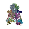
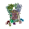
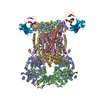
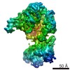
| Title | YEAST CYTOCHROME BC1 COMPLEX WITH BOUND SUBSTRATE CYTOCHROME C | |||||||||
 Components Components |
| |||||||||
 Keywords Keywords | OXIDOREDUCTASE/ELECTRON TRANSPORT / multisubunit membrane protein complex / enzyme substrate complex / electron transfer complex / antibody Fv fragment mediated crystallization / OXIDOREDUCTASE-ELECTRON TRANSPORT COMPLEX | |||||||||
| Function / homology |  Function and homology information Function and homology informationComplex III assembly / : / matrix side of mitochondrial inner membrane / : / Release of apoptotic factors from the mitochondria / Pyroptosis / Detoxification of Reactive Oxygen Species / Respiratory electron transport / Mitochondrial protein degradation / mitochondrial respiratory chain complex III assembly ...Complex III assembly / : / matrix side of mitochondrial inner membrane / : / Release of apoptotic factors from the mitochondria / Pyroptosis / Detoxification of Reactive Oxygen Species / Respiratory electron transport / Mitochondrial protein degradation / mitochondrial respiratory chain complex III assembly / cellular respiration / cardiolipin binding / respiratory chain complex III / quinol-cytochrome-c reductase / mitochondrial electron transport, cytochrome c to oxygen / quinol-cytochrome-c reductase activity / mitochondrial electron transport, ubiquinol to cytochrome c / mitochondrial crista / proton transmembrane transport / nuclear periphery / aerobic respiration / metalloendopeptidase activity / mitochondrial intermembrane space / 2 iron, 2 sulfur cluster binding / electron transfer activity / oxidoreductase activity / mitochondrial inner membrane / heme binding / mitochondrion / proteolysis / metal ion binding / membrane / cytosol Similarity search - Function | |||||||||
| Biological species |   | |||||||||
| Method |  X-RAY DIFFRACTION / SYNCHROTRON / MOLECULAR REPLACEMENT / Resolution: 2.97 Å X-RAY DIFFRACTION / SYNCHROTRON / MOLECULAR REPLACEMENT / Resolution: 2.97 Å | |||||||||
 Authors Authors | Lange, C. / Hunte, C. | |||||||||
 Citation Citation | Journal: Proc.Natl.Acad.Sci.USA / Year: 2002 Title: Crystal structure of the yeast cytochrome bc1 complex with its bound substrate cytochrome c. Authors: Lange, C. / Hunte, C. | |||||||||
| History |
| |||||||||
| Remark 999 | SEQUENCE Residue 270 (chains C and N) is in conflict in swissprot entry P00163 as being either ASP or VAL. |
- Structure visualization
| Structure viewer | Molecule: MolmilJmol/JSmol |
|---|
- Downloads & links
-Download
| PDBx/mmCIF format | 1kyo.cif.gz | 884.3 KB | Display | PDBx/mmCIF format |
|---|---|---|---|---|
| PDB format | pdb1kyo.ent.gz | 717.3 KB | Display | PDB format |
| PDBx/mmJSON format | 1kyo.json.gz | Tree view | PDBx/mmJSON format | |
| Others |  Other downloads Other downloads |
-Validation report
| Arichive directory | https://data.pdbj.org/pub/pdb/validation_reports/ky/1kyoftp://data.pdbj.org/pub/pdb/validation_reports/ky/1kyo | HTTPS FTP |
|---|
-Related structure data
| Related structure data |  1ezvS S: Starting model for refinement |
|---|---|
| Similar structure data |


-Links
 PDBj
PDBj


- Assembly
| Deposited unit | 
| ||||||||
|---|---|---|---|---|---|---|---|---|---|
| 1 |
| ||||||||
| Unit cell |
|
-Components
-UBIQUINOL-CYTOCHROME C REDUCTASE COMPLEX ... , 6 types, 12 molecules ALBMFQGRHSIT
| #1: Protein | Mass: 47358.168 Da / Num. of mol.: 2 / Fragment: RESIDUES 27-457 / Source method: isolated from a natural source / Source: (natural) #2: Protein | Mass: 38751.918 Da / Num. of mol.: 2 / Fragment: RESIDUES 17-368 / Source method: isolated from a natural source / Source: (natural) #6: Protein | Mass: 8870.749 Da / Num. of mol.: 2 / Fragment: RESIDUES 74-147 / Source method: isolated from a natural source / Source: (natural) #7: Protein | Mass: 14452.557 Da / Num. of mol.: 2 / Fragment: RESIDUES 2-127 / Source method: isolated from a natural source / Source: (natural) #8: Protein | Mass: 10856.314 Da / Num. of mol.: 2 / Fragment: RESIDUES 2-94 / Source method: isolated from a natural source / Source: (natural) #9: Protein | Mass: 6507.470 Da / Num. of mol.: 2 / Fragment: RESIDUES 1-57 / Source method: isolated from a natural source / Source: (natural) |
|---|
-Protein , 4 types, 7 molecules CNDOEPW
| #3: Protein | Mass: 43674.535 Da / Num. of mol.: 2 / Source method: isolated from a natural source / Source: (natural) #4: Protein | Mass: 27807.395 Da / Num. of mol.: 2 / Fragment: RESIDUES 62-309 / Source method: isolated from a natural source / Source: (natural) #5: Protein | Mass: 20122.955 Da / Num. of mol.: 2 / Fragment: RESIDUES 31-215 / Source method: isolated from a natural source / Source: (natural) #12: Protein | | Mass: 12115.915 Da / Num. of mol.: 1 / Source method: isolated from a natural source / Source: (natural) |
|---|
-Antibody , 2 types, 4 molecules JUKV
| #10: Antibody | Mass: 14365.817 Da / Num. of mol.: 2 / Fragment: RESIDUES 1-127 Source method: isolated from a genetically manipulated source Source: (gene. exp.)  #11: Antibody | Mass: 11926.350 Da / Num. of mol.: 2 / Fragment: RESIDUES 1-107 Source method: isolated from a genetically manipulated source Source: (gene. exp.) |
|---|
-Non-polymers , 3 types, 11 molecules 

| #13: Chemical | ChemComp-HEC /  Mass: 618.503 Da / Num. of mol.: 7 / Source method: obtained synthetically / Formula: C34H34FeN4O4 Mass: 618.503 Da / Num. of mol.: 7 / Source method: obtained synthetically / Formula: C34H34FeN4O4#14: Chemical |  Mass: 514.650 Da / Num. of mol.: 2 / Source method: obtained synthetically / Formula: C30H42O7 Mass: 514.650 Da / Num. of mol.: 2 / Source method: obtained synthetically / Formula: C30H42O7#15: Chemical |  Mass: 175.820 Da / Num. of mol.: 2 / Source method: obtained synthetically / Formula: Fe2S2 Mass: 175.820 Da / Num. of mol.: 2 / Source method: obtained synthetically / Formula: Fe2S2 |
|---|
-Experimental details
-Experiment
| Experiment | Method: X-RAY DIFFRACTION / Number of used crystals: 1 |
|---|
- Sample preparation
| Crystal | Density Matthews: 4.61 Å3/Da / Density % sol: 73.33 % |
|---|---|
| Crystal grow | Temperature: 277 K / Method: vapor diffusion, hanging drop / pH: 7.5 Details: PEG4000, 20 mM Tris, 0.05 % Undecyl-maltopyranoside, pH 7.5, VAPOR DIFFUSION, HANGING DROP, temperature 277K |
-Data collection
| Diffraction | Mean temperature: 277 K |
|---|---|
| Diffraction source | Source: SYNCHROTRON / Site: ESRF  / Beamline: ID14-3 / Wavelength: 0.931 Å / Beamline: ID14-3 / Wavelength: 0.931 Å |
| Detector | Type: MARRESEARCH / Detector: CCD / Date: Feb 26, 2001 |
| Radiation | Protocol: SINGLE WAVELENGTH / Monochromatic (M) / Laue (L): M / Scattering type: x-ray |
| Radiation wavelength | Wavelength: 0.931 Å / Relative weight: 1 |
| Reflection | Resolution: 2.94→29.64 Å / Num. all: 187312 / Num. obs: 175444 / % possible obs: 92.8 % / Observed criterion σ(F): 0 / Observed criterion σ(I): 0 / Redundancy: 3.2 % / Biso Wilson estimate: 43.7 Å2 / Limit h max: 48 / Limit h min: -49 / Limit k max: 56 / Limit k min: -49 / Limit l max: 66 / Limit l min: 0 / Observed criterion F max: 81215.36 / Observed criterion F min: 0.32 / Rsym value: 0.106 / Net I/σ(I): 6.8 |
| Reflection shell | Resolution: 2.97→3.08 Å / Mean I/σ(I) obs: 2.3 / Num. unique all: 14711 / Rsym value: 0.395 / % possible all: 76.5 |
| Reflection | *PLUS Highest resolution: 2.97 Å / Lowest resolution: 30 Å / Rmerge(I) obs: 0.106 |
| Reflection shell | *PLUS % possible obs: 76.5 % |
- Processing
| Software |
| ||||||||||||||||||||||||||||||||||||||||||||||||||||||||||||||||||||||||||||||||||||||||||
|---|---|---|---|---|---|---|---|---|---|---|---|---|---|---|---|---|---|---|---|---|---|---|---|---|---|---|---|---|---|---|---|---|---|---|---|---|---|---|---|---|---|---|---|---|---|---|---|---|---|---|---|---|---|---|---|---|---|---|---|---|---|---|---|---|---|---|---|---|---|---|---|---|---|---|---|---|---|---|---|---|---|---|---|---|---|---|---|---|---|---|---|
| Refinement | Method to determine structure: MOLECULAR REPLACEMENT Starting model: PDB ENTRY 1EZV Resolution: 2.97→29.64 Å / Rfactor Rfree error: 0.002 / Occupancy max: 1 / Occupancy min: 1 / Cross valid method: THROUGHOUT / σ(F): 0 / Stereochemistry target values: Engh & Huber
| ||||||||||||||||||||||||||||||||||||||||||||||||||||||||||||||||||||||||||||||||||||||||||
| Solvent computation | Solvent model: CNS bulk solvent model used / Bsol: 10 Å2 / ksol: 0.228013 e/Å3 | ||||||||||||||||||||||||||||||||||||||||||||||||||||||||||||||||||||||||||||||||||||||||||
| Displacement parameters | Biso max: 162.05 Å2 / Biso mean: 59.66 Å2 / Biso min: 0.22 Å2
| ||||||||||||||||||||||||||||||||||||||||||||||||||||||||||||||||||||||||||||||||||||||||||
| Refine analyze |
| ||||||||||||||||||||||||||||||||||||||||||||||||||||||||||||||||||||||||||||||||||||||||||
| Refinement step | Cycle: LAST / Resolution: 2.97→29.64 Å
| ||||||||||||||||||||||||||||||||||||||||||||||||||||||||||||||||||||||||||||||||||||||||||
| Refine LS restraints |
| ||||||||||||||||||||||||||||||||||||||||||||||||||||||||||||||||||||||||||||||||||||||||||
| LS refinement shell | Refine-ID: X-RAY DIFFRACTION / Total num. of bins used: 8
| ||||||||||||||||||||||||||||||||||||||||||||||||||||||||||||||||||||||||||||||||||||||||||
| Software | *PLUS Name: CNS / Version: 1 / Classification: refinement | ||||||||||||||||||||||||||||||||||||||||||||||||||||||||||||||||||||||||||||||||||||||||||
| Refinement | *PLUS σ(F): 0 / % reflection Rfree: 9.3 % / Rfactor obs: 0.228 / Rfactor Rfree: 0.267 | ||||||||||||||||||||||||||||||||||||||||||||||||||||||||||||||||||||||||||||||||||||||||||
| Solvent computation | *PLUS | ||||||||||||||||||||||||||||||||||||||||||||||||||||||||||||||||||||||||||||||||||||||||||
| Displacement parameters | *PLUS | ||||||||||||||||||||||||||||||||||||||||||||||||||||||||||||||||||||||||||||||||||||||||||
| Refine LS restraints | *PLUS
| ||||||||||||||||||||||||||||||||||||||||||||||||||||||||||||||||||||||||||||||||||||||||||
| LS refinement shell | *PLUS Rfactor Rfree: 0.393 / % reflection Rfree: 8.5 % / Rfactor Rwork: 0.393 |
