 Movie
Movie Controller


+ Open data

- Basic information
| Entry | Database: PDB / ID: 1kp4 | ||||||
|---|---|---|---|---|---|---|---|
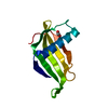
| Title | CALCIUM-BOUND FORM OF PROKARYOTIC PHOSPHOLIPASE A2 | ||||||
 Components Components | phospholipase A2 | ||||||
 Keywords Keywords | HYDROLASE / Phospholipase A2 / Prokaryote / calcium ion | ||||||
| Function / homology |  Function and homology information Function and homology informationA2-type glycerophospholipase activity / arachidonate secretion / phospholipid metabolic process / metal ion binding Similarity search - Function | ||||||
| Biological species |  Streptomyces violaceoruber (bacteria) Streptomyces violaceoruber (bacteria) | ||||||
| Method |  X-RAY DIFFRACTION / AB INITIO / Resolution: 1.6 Å X-RAY DIFFRACTION / AB INITIO / Resolution: 1.6 Å | ||||||
 Authors Authors | Matoba, Y. / Katsube, Y. / Sugiyama, M. | ||||||
 Citation Citation | Journal: J.Biol.Chem. / Year: 2002 Title: The crystal structure of prokaryotic phospholipase A2. Authors: Matoba, Y. / Katsube, Y. / Sugiyama, M. #1: Journal: J.Biol.Chem. / Year: 2002Title: A novel prokaryotic phospholipase A2. Characterization, gene cloning, and solution structure Authors: Sugiyama, M. / Ohtani, K. / Izuhara, M. / Koike, T. / Suzuki, K. / Imamura, S. / Misaki, H. | ||||||
| History |
|
- Structure visualization
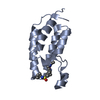
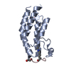
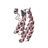
| Structure viewer | Molecule: MolmilJmol/JSmol |
|---|
- Downloads & links
-Download
| PDBx/mmCIF format | 1kp4.cif.gz | 38.5 KB | Display | PDBx/mmCIF format |
|---|---|---|---|---|
| PDB format | pdb1kp4.ent.gz | 25.6 KB | Display | PDB format |
| PDBx/mmJSON format | 1kp4.json.gz | Tree view | PDBx/mmJSON format | |
| Others |  Other downloads Other downloads |
-Validation report
| Arichive directory | https://data.pdbj.org/pub/pdb/validation_reports/kp/1kp4ftp://data.pdbj.org/pub/pdb/validation_reports/kp/1kp4 | HTTPS FTP |
|---|
-Related structure data
| Related structure data |  1fazSC S: Starting model for refinement C: citing same article ( |
|---|---|
| Similar structure data |


-Links
 PDBj
PDBj
- Assembly
| Deposited unit | 
| ||||||||
|---|---|---|---|---|---|---|---|---|---|
| 1 |
| ||||||||
| Unit cell |
| ||||||||
| Details | The biological unit of this protein is a monomer |
-Components
| #1: Protein | Mass: 13570.782 Da / Num. of mol.: 1 Source method: isolated from a genetically manipulated source Source: (gene. exp.) Streptomyces violaceoruber (bacteria) / Plasmid: pIJ680 / Species (production host): Streptomyces lividans / Production host: Streptomyces lividans TK24 (bacteria) / Strain (production host): TK24References: UniProt: Q9Z4W2, UniProt: Q6UV28*PLUS, phospholipase A2 |
|---|---|
| #2: Chemical | ChemComp-CA /   Mass: 40.078 Da / Num. of mol.: 1 / Source method: obtained synthetically / Formula: Ca Mass: 40.078 Da / Num. of mol.: 1 / Source method: obtained synthetically / Formula: Ca |
| #3: Water | ChemComp-HOH /  Mass: 18.015 Da / Num. of mol.: 88 / Source method: isolated from a natural source / Formula: H2O Mass: 18.015 Da / Num. of mol.: 88 / Source method: isolated from a natural source / Formula: H2O |
| Has protein modification | Y |
-Experimental details
-Experiment
| Experiment | Method: X-RAY DIFFRACTION / Number of used crystals: 1 |
|---|
- Sample preparation
| Crystal | Density Matthews: 2.02 Å3/Da / Density % sol: 47.53 % |
|---|---|
| Crystal grow | Temperature: 297 K / Method: vapor diffusion, hanging drop / pH: 8.5 Details: MPD, Tris-HCl, pH 8.5, VAPOR DIFFUSION, HANGING DROP, temperature 297K |
-Data collection
| Diffraction | Mean temperature: 292 K |
|---|---|
| Diffraction source | Source: ROTATING ANODE / Type: RIGAKU RU300 / Wavelength: 1.5418 Å |
| Detector | Type: RIGAKU RAXIS IIC / Detector: IMAGE PLATE / Date: Mar 1, 1999 / Details: mirrors |
| Radiation | Monochromator: MIRRORS / Protocol: SINGLE WAVELENGTH / Monochromatic (M) / Laue (L): M / Scattering type: x-ray |
| Radiation wavelength | Wavelength: 1.5418 Å / Relative weight: 1 |
| Reflection | Resolution: 1.6→100 Å / Num. all: 14730 / Num. obs: 12820 / % possible obs: 73.2 % / Observed criterion σ(F): 2 / Observed criterion σ(I): 1 / Redundancy: 2.37 % / Rmerge(I) obs: 0.065 / Net I/σ(I): 11.2 |
| Reflection shell | Resolution: 1.6→1.7 Å / Rmerge(I) obs: 0.283 / Mean I/σ(I) obs: 2.41 / Num. unique all: 1325 / % possible all: 48 |
- Processing
| Software |
| |||||||||||||||||||||||||||||||||
|---|---|---|---|---|---|---|---|---|---|---|---|---|---|---|---|---|---|---|---|---|---|---|---|---|---|---|---|---|---|---|---|---|---|---|
| Refinement | Method to determine structure: AB INITIO Starting model: PDB ENTRY 1FAZ Resolution: 1.6→5 Å / Num. parameters: 4191 / Num. restraintsaints: 3988 / Isotropic thermal model: isotropic / σ(F): 0 / Stereochemistry target values: ENGH AND HUBER
| |||||||||||||||||||||||||||||||||
| Refine analyze | Luzzati coordinate error obs: 0.16 Å / Luzzati d res low obs: 5 Å / Num. disordered residues: 0 / Occupancy sum hydrogen: 0 / Occupancy sum non hydrogen: 1047 | |||||||||||||||||||||||||||||||||
| Refinement step | Cycle: LAST / Resolution: 1.6→5 Å
| |||||||||||||||||||||||||||||||||
| Refine LS restraints |
| |||||||||||||||||||||||||||||||||
| LS refinement shell | Resolution: 1.6→1.7 Å
|
