 Movie
Movie Controller

[English] 日本語
 Yorodumi
- PDB-5u9n: Second Bromodomain of cdg4_1340 from Cryptosporidium parvum, comp... -
+ Open data

- Basic information
| Entry | Database: PDB / ID: 5u9n | ||||||
|---|---|---|---|---|---|---|---|
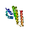
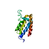
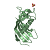
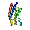
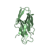
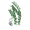
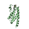
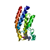
| Title | Second Bromodomain of cdg4_1340 from Cryptosporidium parvum, complexed with bromosporine | ||||||
 Components Components | Bromo domain containing protein | ||||||
 Keywords Keywords | SIGNALING PROTEIN / Bromodomain / Ligand / Structural Genomics Consortium (SGC) | ||||||
| Function / homology |  Function and homology information Function and homology informationBromodomain-like / Histone Acetyltransferase; Chain A / Bromodomain, conserved site / Bromodomain signature. / Bromodomain / bromo domain / Bromodomain / Bromodomain (BrD) profile. / Bromodomain-like superfamily / Up-down Bundle / Mainly Alpha Similarity search - Domain/homology | ||||||
| Biological species |   Cryptosporidium parvum (eukaryote) Cryptosporidium parvum (eukaryote) | ||||||
| Method |  X-RAY DIFFRACTION / SYNCHROTRON / MOLECULAR REPLACEMENT / molecular replacement / Resolution: 2.4 Å X-RAY DIFFRACTION / SYNCHROTRON / MOLECULAR REPLACEMENT / molecular replacement / Resolution: 2.4 Å | ||||||
| Model details | In subunit B the protein appears to be significantly disordered, and while some residues in the ...In subunit B the protein appears to be significantly disordered, and while some residues in the range 413-420 have been placed into the density, there is a possibility that the docking is incorrect. The user should use caution in interpreting these results. | ||||||
 Authors Authors | Hou, C.F.D. / Lin, Y.H. / Loppnau, P. / Hutchinson, A. / Dong, A. / Bountra, C. / Edwards, A.M. / Arrowsmith, C.H. / Hui, R. / Walker, J.R. / Structural Genomics Consortium (SGC) | ||||||
 Citation Citation | Journal: To be published Title: Second Bromodomain of cdg4_1340 from Cryptosporidium parvum, complexed with bromosporine Authors: Hou, C.F.D. / Lin, Y.H. / Loppnau, P. / Hutchinson, A. / Dong, A. / Bountra, C. / Edwards, A.M. / Arrowsmith, C.H. / Hui, R. / Walker, J.R. / Structural Genomics Consortium (SGC) | ||||||
| History |
|
- Structure visualization
| Structure viewer | Molecule: MolmilJmol/JSmol |
|---|
- Downloads & links
-Download
| PDBx/mmCIF format | 5u9n.cif.gz | 120.9 KB | Display | PDBx/mmCIF format |
|---|---|---|---|---|
| PDB format | pdb5u9n.ent.gz | 92.9 KB | Display | PDB format |
| PDBx/mmJSON format | 5u9n.json.gz | Tree view | PDBx/mmJSON format | |
| Others |  Other downloads Other downloads |
-Validation report
| Arichive directory | https://data.pdbj.org/pub/pdb/validation_reports/u9/5u9nftp://data.pdbj.org/pub/pdb/validation_reports/u9/5u9n | HTTPS FTP |
|---|
-Related structure data
| Related structure data | 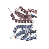 4py6S S: Starting model for refinement |
|---|---|
| Similar structure data |


-Links
 PDBj
PDBj- Assembly
| Deposited unit | 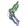
| ||||||||
|---|---|---|---|---|---|---|---|---|---|
| 1 | 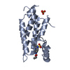
| ||||||||
| 2 | 
| ||||||||
| Unit cell |
|
-Components
| #1: Protein | Mass: 17863.914 Da / Num. of mol.: 2 / Fragment: Second bromodomain (UNP residues 300-450) Source method: isolated from a genetically manipulated source Source: (gene. exp.) Cryptosporidium parvum (strain Iowa II) (eukaryote)Strain: Iowa II / Gene: cgd4_1340 / Plasmid: PET15-MHL / Production host:  #2: Chemical |   Mass: 404.444 Da / Num. of mol.: 2 / Source method: obtained synthetically / Formula: C17H20N6O4S Mass: 404.444 Da / Num. of mol.: 2 / Source method: obtained synthetically / Formula: C17H20N6O4S#3: Chemical | ChemComp-SO4 / |   Mass: 96.063 Da / Num. of mol.: 1 / Source method: obtained synthetically / Formula: SO4 Mass: 96.063 Da / Num. of mol.: 1 / Source method: obtained synthetically / Formula: SO4#4: Water | ChemComp-HOH / |  Mass: 18.015 Da / Num. of mol.: 75 / Source method: isolated from a natural source / Formula: H2O Mass: 18.015 Da / Num. of mol.: 75 / Source method: isolated from a natural source / Formula: H2O |
|---|
-Experimental details
-Experiment
| Experiment | Method: X-RAY DIFFRACTION / Number of used crystals: 1 |
|---|
- Sample preparation
| Crystal | Density Matthews: 3.95 Å3/Da / Density % sol: 68.84 % |
|---|---|
| Crystal grow | Temperature: 293 K / Method: vapor diffusion, sitting drop / pH: 7 Details: The protein was crystallized at 293 K in 2.5M ammonium sulfate, 0.1 M bis-tris propane pH 7.0. Bromosporine (ethyl (3-methyl-6-{4-methyl-3-[(methylsulfonyl)amino]phenyl}[1,2,4]triazolo[4,3- ...Details: The protein was crystallized at 293 K in 2.5M ammonium sulfate, 0.1 M bis-tris propane pH 7.0. Bromosporine (ethyl (3-methyl-6-{4-methyl-3-[(methylsulfonyl)amino]phenyl}[1,2,4]triazolo[4,3- b]pyridazin-8-yl)carbamate) was added (final concentration of 1 mM)directly to the concentrated protein immediately prior to setting up the crystallization plate |
-Data collection
| Diffraction | Mean temperature: 100 K | ||||||||||||||||||
|---|---|---|---|---|---|---|---|---|---|---|---|---|---|---|---|---|---|---|---|
| Diffraction source | Source: SYNCHROTRON / Site: APS  / Beamline: 22-ID / Wavelength: 0.976 Å / Beamline: 22-ID / Wavelength: 0.976 Å | ||||||||||||||||||
| Detector | Type: MARMOSAIC 300 mm CCD / Detector: CCD / Date: Nov 25, 2016 | ||||||||||||||||||
| Radiation | Protocol: SINGLE WAVELENGTH / Monochromatic (M) / Laue (L): M / Scattering type: x-ray | ||||||||||||||||||
| Radiation wavelength | Wavelength: 0.976 Å / Relative weight: 1 | ||||||||||||||||||
| Reflection | Resolution: 2.4→33.67 Å / Num. obs: 22425 / % possible obs: 99.8 % / Redundancy: 7.1 % / Biso Wilson estimate: 57.95 Å2 / CC1/2: 0.996 / Rmerge(I) obs: 0.1 / Rpim(I) all: 0.041 / Rrim(I) all: 0.108 / Net I/σ(I): 18 / Num. measured all: 159515 / Scaling rejects: 1179 | ||||||||||||||||||
| Reflection shell |
|
-Phasing
| Phasing | Method: molecular replacement |
|---|
- Processing
| Software |
| ||||||||||||||||||||||||||||||||||||||||||||||||||||||||||||||||||||||||||||||||||||||||||||||||||||||||||||
|---|---|---|---|---|---|---|---|---|---|---|---|---|---|---|---|---|---|---|---|---|---|---|---|---|---|---|---|---|---|---|---|---|---|---|---|---|---|---|---|---|---|---|---|---|---|---|---|---|---|---|---|---|---|---|---|---|---|---|---|---|---|---|---|---|---|---|---|---|---|---|---|---|---|---|---|---|---|---|---|---|---|---|---|---|---|---|---|---|---|---|---|---|---|---|---|---|---|---|---|---|---|---|---|---|---|---|---|---|---|
| Refinement | Method to determine structure: MOLECULAR REPLACEMENT Starting model: 4PY6 Resolution: 2.4→32.54 Å / Cor.coef. Fo:Fc: 0.913 / Cor.coef. Fo:Fc free: 0.884 / SU R Cruickshank DPI: 0.304 / Cross valid method: THROUGHOUT / σ(F): 0 / SU R Blow DPI: 0.209 / SU Rfree Blow DPI: 0.187 / SU Rfree Cruickshank DPI: 0.188 Details: In subunit B the protein appears to be significantly disordered, and while some residues in the range 413-420 have been placed into the density, there is a possibility that the docking is ...Details: In subunit B the protein appears to be significantly disordered, and while some residues in the range 413-420 have been placed into the density, there is a possibility that the docking is incorrect. The user should use caution in interpreting these results.
| ||||||||||||||||||||||||||||||||||||||||||||||||||||||||||||||||||||||||||||||||||||||||||||||||||||||||||||
| Displacement parameters | Biso max: 159.17 Å2 / Biso mean: 67.55 Å2 / Biso min: 26.63 Å2
| ||||||||||||||||||||||||||||||||||||||||||||||||||||||||||||||||||||||||||||||||||||||||||||||||||||||||||||
| Refine analyze | Luzzati coordinate error obs: 0.38 Å | ||||||||||||||||||||||||||||||||||||||||||||||||||||||||||||||||||||||||||||||||||||||||||||||||||||||||||||
| Refinement step | Cycle: final / Resolution: 2.4→32.54 Å
| ||||||||||||||||||||||||||||||||||||||||||||||||||||||||||||||||||||||||||||||||||||||||||||||||||||||||||||
| Refine LS restraints |
| ||||||||||||||||||||||||||||||||||||||||||||||||||||||||||||||||||||||||||||||||||||||||||||||||||||||||||||
| LS refinement shell | Resolution: 2.4→2.52 Å / Rfactor Rfree error: 0 / Total num. of bins used: 11
| ||||||||||||||||||||||||||||||||||||||||||||||||||||||||||||||||||||||||||||||||||||||||||||||||||||||||||||
| Refinement TLS params. | Method: refined / Refine-ID: X-RAY DIFFRACTION
| ||||||||||||||||||||||||||||||||||||||||||||||||||||||||||||||||||||||||||||||||||||||||||||||||||||||||||||
| Refinement TLS group |
|
