 Movie
Movie Controller


+ Open data

- Basic information
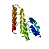
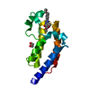
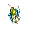
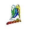
| Entry | Database: PDB / ID: 1faz | ||||||
|---|---|---|---|---|---|---|---|
| Title | THE CRYSTAL STRUCTURE OF PROKARYOTIC PHOSPHOLIPASE A2 | ||||||
 Components Components | PHOSPHOLIPASE A2 | ||||||
 Keywords Keywords | HYDROLASE / phospholipase A2 / prokaryote / Streptomyces violaceoruber | ||||||
| Function / homology |  Function and homology information Function and homology informationA2-type glycerophospholipase activity / arachidonate secretion / phospholipid metabolic process / metal ion binding Similarity search - Function | ||||||
| Biological species |  Streptomyces violaceoruber (bacteria) Streptomyces violaceoruber (bacteria) | ||||||
| Method |  X-RAY DIFFRACTION / Resolution: 1.4 Å X-RAY DIFFRACTION / Resolution: 1.4 Å | ||||||
 Authors Authors | Matoba, Y. / katsube, Y. / Sugiyama, M. | ||||||
 Citation Citation | Journal: J.Biol.Chem. / Year: 2002 Title: The crystal structure of prokaryotic phospholipase A2. Authors: Matoba, Y. / Katsube, Y. / Sugiyama, M. | ||||||
| History |
|
- Structure visualization
| Structure viewer | Molecule: MolmilJmol/JSmol |
|---|
- Downloads & links
-Download
| PDBx/mmCIF format | 1faz.cif.gz | 37.6 KB | Display | PDBx/mmCIF format |
|---|---|---|---|---|
| PDB format | pdb1faz.ent.gz | 25.1 KB | Display | PDB format |
| PDBx/mmJSON format | 1faz.json.gz | Tree view | PDBx/mmJSON format | |
| Others |  Other downloads Other downloads |
-Validation report
| Arichive directory | https://data.pdbj.org/pub/pdb/validation_reports/fa/1fazftp://data.pdbj.org/pub/pdb/validation_reports/fa/1faz | HTTPS FTP |
|---|
-Related structure data


-Links
 PDBj
PDBj
- Assembly
| Deposited unit | 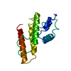
| ||||||||
|---|---|---|---|---|---|---|---|---|---|
| 1 |
| ||||||||
| Unit cell |
|
-Components
| #1: Protein | Mass: 13570.782 Da / Num. of mol.: 1 Source method: isolated from a genetically manipulated source Source: (gene. exp.) Streptomyces violaceoruber (bacteria) / Description: SECRETED / Plasmid: PIJ680 / Production host: Streptomyces lividans (bacteria)References: UniProt: Q9Z4W2, UniProt: Q6UV28*PLUS, phospholipase A2 |
|---|---|
| #2: Water | ChemComp-HOH /  Mass: 18.015 Da / Num. of mol.: 127 / Source method: isolated from a natural source / Formula: H2O Mass: 18.015 Da / Num. of mol.: 127 / Source method: isolated from a natural source / Formula: H2O |
| Has protein modification | Y |
-Experimental details
-Experiment
| Experiment | Method: X-RAY DIFFRACTION / Number of used crystals: 1 |
|---|
- Sample preparation
| Crystal | Density Matthews: 1.85 Å3/Da / Density % sol: 33.36 % | ||||||||||||||||||||||||||||||||||||||||||
|---|---|---|---|---|---|---|---|---|---|---|---|---|---|---|---|---|---|---|---|---|---|---|---|---|---|---|---|---|---|---|---|---|---|---|---|---|---|---|---|---|---|---|---|
| Crystal grow | Temperature: 298 K / Method: vapor diffusion, hanging drop / pH: 6 Details: PEG 4000, lithium sulfate, sodium cacodylate, pH 6, VAPOR DIFFUSION, HANGING DROP, temperature 298K | ||||||||||||||||||||||||||||||||||||||||||
| Crystal grow | *PLUS Temperature: 297 K / pH: 8 | ||||||||||||||||||||||||||||||||||||||||||
| Components of the solutions | *PLUS
|
-Data collection
| Diffraction | Mean temperature: 298 K |
|---|---|
| Diffraction source | Source: ROTATING ANODE / Type: RIGAKU RU200 / Wavelength: 1.5418 |
| Detector | Type: RIGAKU RAXIS IIC / Detector: IMAGE PLATE |
| Radiation | Protocol: SINGLE WAVELENGTH / Monochromatic (M) / Laue (L): M / Scattering type: x-ray |
| Radiation wavelength | Wavelength: 1.5418 Å / Relative weight: 1 |
| Reflection | Resolution: 1.4→100 Å / Num. all: 16049 / Num. obs: 15558 / % possible obs: 77.5 % / Observed criterion σ(F): 2 / Observed criterion σ(I): 1 / Redundancy: 1.41 % / Biso Wilson estimate: 11.7 Å2 / Rmerge(I) obs: 0.074 / Net I/σ(I): 34.2 |
| Reflection shell | Resolution: 1.4→1.45 Å / Redundancy: 1.4 % / Rmerge(I) obs: 0.276 / Num. unique all: 1174 / % possible all: 61.1 |
| Reflection | *PLUS Lowest resolution: 100 Å / Num. measured all: 22015 / Rmerge(I) obs: 0.0741 |
- Processing
| Software |
| ||||||||||||||||||||||||||||||||||||||||
|---|---|---|---|---|---|---|---|---|---|---|---|---|---|---|---|---|---|---|---|---|---|---|---|---|---|---|---|---|---|---|---|---|---|---|---|---|---|---|---|---|---|
| Refinement | Resolution: 1.4→10 Å / Rfactor Rfree error: 0.006 / Data cutoff high absF: 10000000 / Data cutoff low absF: 0 / Isotropic thermal model: RESTRAINED / Cross valid method: THROUGHOUT / σ(F): 2 / σ(I): 1 / Stereochemistry target values: Engh & Huber
| ||||||||||||||||||||||||||||||||||||||||
| Displacement parameters | Biso mean: 13.1 Å2
| ||||||||||||||||||||||||||||||||||||||||
| Refine analyze |
| ||||||||||||||||||||||||||||||||||||||||
| Refinement step | Cycle: LAST / Resolution: 1.4→10 Å
| ||||||||||||||||||||||||||||||||||||||||
| Refine LS restraints |
| ||||||||||||||||||||||||||||||||||||||||
| LS refinement shell | Resolution: 1.4→1.49 Å / Rfactor Rfree error: 0.018 / Total num. of bins used: 6
| ||||||||||||||||||||||||||||||||||||||||
| Xplor file |
| ||||||||||||||||||||||||||||||||||||||||
| Software | *PLUS Name: X-PLOR / Version: 3.851 / Classification: refinement | ||||||||||||||||||||||||||||||||||||||||
| Refinement | *PLUS σ(F): 2 / % reflection Rfree: 9.8 % | ||||||||||||||||||||||||||||||||||||||||
| Solvent computation | *PLUS | ||||||||||||||||||||||||||||||||||||||||
| Displacement parameters | *PLUS Biso mean: 13.1 Å2 | ||||||||||||||||||||||||||||||||||||||||
| Refine LS restraints | *PLUS
| ||||||||||||||||||||||||||||||||||||||||
| LS refinement shell | *PLUS Rfactor Rfree: 0.244 / % reflection Rfree: 9.8 % / Rfactor Rwork: 0.239 |
