[English] 日本語
Yorodumi
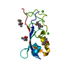
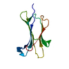
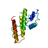
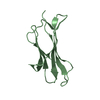
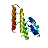
- PDB-6t3o: Crystal structure of the human myomesin domain 10 -

+
Open data


ID or keywords:

Loading...

-
Basic information

Entry
Database: PDB / ID: 6t3o
TitleCrystal structure of the human myomesin domain 10
ComponentsMyomesin-1
KeywordsCONTRACTILE PROTEIN / small protein domain / scaffold structure
Function / homology
Function and homology information


extraocular skeletal muscle development / striated muscle myosin thick filament / M band / structural constituent of muscle / sarcomere organization / protein kinase A signaling / positive regulation of protein secretion / kinase binding / positive regulation of gene expression / protein homodimerization activity / identical protein binding
Similarity search - Function
Immunoglobulin I-set / Immunoglobulin I-set domain / Fibronectin type III domain / Fibronectin type 3 domain / Immunoglobulin subtype 2 / Immunoglobulin C-2 Type / Fibronectin type-III domain profile. / Fibronectin type III / Fibronectin type III superfamily / Immunoglobulin subtype ...Immunoglobulin I-set / Immunoglobulin I-set domain / Fibronectin type III domain / Fibronectin type 3 domain / Immunoglobulin subtype 2 / Immunoglobulin C-2 Type / Fibronectin type-III domain profile. / Fibronectin type III / Fibronectin type III superfamily / Immunoglobulin subtype / Immunoglobulin / Ig-like domain profile. / Immunoglobulin-like domain / Immunoglobulin-like domain superfamily / Immunoglobulin-like fold
Similarity search - Domain/homology
AZIDE ION / Myomesin-1
Similarity search - Component
Biological speciesHomo sapiens (human)
MethodX-RAY DIFFRACTION / SYNCHROTRON / MOLECULAR REPLACEMENT / Resolution: 1.8 Å
AuthorsDuskova, J. / Petrokova, H. / Maly, P.
Funding support Czech Republic, 1items
OrganizationGrant numberCountry
Ministry of Education, Youth and Sports of the Czech RepublicCZ.02.1.01/0.0/0.0/16_025/0007397 Czech Republic
CitationJournal: Virulence / Year: 2021
Title: Myomedin scaffold variants targeted to 10E8 HIV-1 broadly neutralizing antibody mimic gp41 epitope and elicit HIV-1 virus-neutralizing sera in mice.
Authors: Kuchar, M. / Kosztyu, P. / Daniel Liskova, V. / Cerny, J. / Petrokova, H. / Vroblova, E. / Maly, M. / Vankova, L. / Krupka, M. / Raskova Kafkova, L. / Turanek Knotigova, P. / Duskova, J. / ...Authors: Kuchar, M. / Kosztyu, P. / Daniel Liskova, V. / Cerny, J. / Petrokova, H. / Vroblova, E. / Maly, M. / Vankova, L. / Krupka, M. / Raskova Kafkova, L. / Turanek Knotigova, P. / Duskova, J. / Dohnalek, J. / Masek, J. / Turanek, J. / Raska, M. / Maly, P.
History
DepositionOct 11, 2019Deposition site: PDBE / Processing site: PDBE
Revision 1.0Mar 17, 2021Provider: repository / Type: Initial release
Revision 1.1Sep 29, 2021Group: Data collection / Database references / Structure summary
Category: citation / citation_author ...citation / citation_author / database_2 / pdbx_database_proc / struct
Item: _citation.journal_abbrev / _citation.journal_id_CSD ..._citation.journal_abbrev / _citation.journal_id_CSD / _citation.journal_id_ISSN / _citation.journal_volume / _citation.page_first / _citation.page_last / _citation.pdbx_database_id_DOI / _citation.pdbx_database_id_PubMed / _citation.title / _citation.year / _database_2.pdbx_DOI / _database_2.pdbx_database_accession / _struct.title
Revision 1.2Jan 24, 2024Group: Data collection / Refinement description
Category: chem_comp_atom / chem_comp_bond / pdbx_initial_refinement_model

-
Structure visualization

Structure viewerMolecule:
MolmilJmol/JSmol

Downloads & links

-
Assembly

Deposited unit
A: Myomesin-1
hetero molecules


Theoretical massNumber of molelcules
Total (without water)15,4372
Polymers15,3951
Non-polymers421
Water1,56787
1


  • Idetical with deposited unit
  • defined by author&software
  • Evidence: gel filtration
TypeNameSymmetry operationNumber
identity operation1_555x,y,z1
Buried area120 Å2
ΔGint1 kcal/mol
Surface area6670 Å2
MethodPISA
Unit cell
Length a, b, c (Å)58.599, 58.599, 62.821
Angle α, β, γ (deg.)90.00, 90.00, 90.00
Int Tables number96
Space group name H-MP43212
Components on special symmetry positions
IDModelComponents
11A-201-

AZI

-
Components

#1: Protein Myomesin-1 / 190 kDa connectin-associated protein / 190 kDa titin-associated protein / Myomesin family member 1


Mass: 15395.444 Da / Num. of mol.: 1
Source method: isolated from a genetically manipulated source
Source: (gene. exp.) Homo sapiens (human) / Gene: MYOM1 / Production host: Escherichia coli (E. coli) / References: UniProt: P52179
#2: Chemical ChemComp-AZI / AZIDE ION


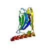
Mass: 42.020 Da / Num. of mol.: 1 / Source method: obtained synthetically / Formula: N3
#3: Water ChemComp-HOH / water


Mass: 18.015 Da / Num. of mol.: 87 / Source method: isolated from a natural source / Formula: H2O
Has ligand of interestN

-
Experimental details

-
Experiment

ExperimentMethod: X-RAY DIFFRACTION / Number of used crystals: 1

-
Sample preparation

CrystalDensity Matthews: 2.09 Å3/Da / Density % sol: 41.12 %
Crystal growTemperature: 291 K / Method: vapor diffusion, hanging drop / pH: 5.4
Details: 0.1 M citrate/phosphate buffer pH 5.4, 30 % PEG3350

-
Data collection

DiffractionMean temperature: 100 K / Serial crystal experiment: N
Diffraction sourceSource: SYNCHROTRON / Site: BESSY / Beamline: 14.1 / Wavelength: 0.918409 Å
DetectorType: DECTRIS PILATUS 300K / Detector: PIXEL / Date: May 30, 2013
RadiationProtocol: SINGLE WAVELENGTH / Monochromatic (M) / Laue (L): M / Scattering type: x-ray
Radiation wavelengthWavelength: 0.918409 Å / Relative weight: 1
ReflectionResolution: 1.8→42.85 Å / Num. all: 55419 / Num. obs: 10591 / % possible obs: 99.7 % / Redundancy: 5.2 % / Biso Wilson estimate: 30.9 Å2 / CC1/2: 1 / Rmerge(I) obs: 0.025 / Rpim(I) all: 0.017 / Rrim(I) all: 0.031 / Χ2: 1 / Net I/av σ(I): 2.5 / Net I/σ(I): 2.5
Reflection shellResolution: 1.8→1.84 Å / Redundancy: 5.4 % / Rmerge(I) obs: 0.572 / Mean I/σ(I) obs: 2.5 / Num. unique obs: 3310 / CC1/2: 0.829 / Rpim(I) all: 0.408 / Rrim(I) all: 0.706 / Χ2: 0.99 / % possible all: 99.9

-
Processing

Software
NameVersionClassification
REFMAC5.8.0238refinement
PDB_EXTRACT3.25data extraction
XDSdata reduction
SCALEPACKdata scaling
MOLREPphasing
RefinementMethod to determine structure: MOLECULAR REPLACEMENT
Starting model: 2Y23
Resolution: 1.8→42.85 Å / Cor.coef. Fo:Fc: 0.955 / SU B: 5.418 / SU ML: 0.086 / Cross valid method: FREE R-VALUE / ESU R: 0.145 / Details: HYDROGENS HAVE BEEN ADDED IN THE RIDING POSITIONS
RfactorNum. reflection% reflection
Rfree0.244 -5 %
Rwork0.1983 --
obs0.20471 10572 99.33 %
Solvent computationIon probe radii: 0.8 Å / Shrinkage radii: 0.8 Å / VDW probe radii: 1.2 Å
Displacement parametersBiso mean: 38.438 Å2
Baniso -1Baniso -2Baniso -3
1--0.04 Å20 Å20 Å2
2---0.04 Å20 Å2
3---0.08 Å2
Refinement stepCycle: LAST / Resolution: 1.8→42.85 Å
ProteinNucleic acidLigandSolventTotal
Num. atoms908 0 3 87 998
Refine LS restraints
Refine-IDTypeDev idealDev ideal targetNumber
X-RAY DIFFRACTIONr_bond_refined_d0.0180.013932
X-RAY DIFFRACTIONr_bond_other_d0.0020.018873
X-RAY DIFFRACTIONr_angle_refined_deg1.9951.6481246
X-RAY DIFFRACTIONr_angle_other_deg1.5211.5872037
X-RAY DIFFRACTIONr_dihedral_angle_1_deg7.5475110
X-RAY DIFFRACTIONr_dihedral_angle_2_deg34.96824.44454
X-RAY DIFFRACTIONr_dihedral_angle_3_deg18.21615185
X-RAY DIFFRACTIONr_dihedral_angle_4_deg23.221154
X-RAY DIFFRACTIONr_chiral_restr0.0930.2116
X-RAY DIFFRACTIONr_gen_planes_refined0.0120.021029
X-RAY DIFFRACTIONr_gen_planes_other0.0020.02191
X-RAY DIFFRACTIONr_nbd_refined
X-RAY DIFFRACTIONr_nbd_other
X-RAY DIFFRACTIONr_nbtor_refined
X-RAY DIFFRACTIONr_nbtor_other
X-RAY DIFFRACTIONr_xyhbond_nbd_refined
X-RAY DIFFRACTIONr_xyhbond_nbd_other
X-RAY DIFFRACTIONr_metal_ion_refined
X-RAY DIFFRACTIONr_metal_ion_other
X-RAY DIFFRACTIONr_symmetry_vdw_refined
X-RAY DIFFRACTIONr_symmetry_vdw_other
X-RAY DIFFRACTIONr_symmetry_hbond_refined
X-RAY DIFFRACTIONr_symmetry_hbond_other
X-RAY DIFFRACTIONr_symmetry_metal_ion_refined
X-RAY DIFFRACTIONr_symmetry_metal_ion_other
X-RAY DIFFRACTIONr_mcbond_it1.8072.451437
X-RAY DIFFRACTIONr_mcbond_other1.8022.447436
X-RAY DIFFRACTIONr_mcangle_it2.473.672545
X-RAY DIFFRACTIONr_mcangle_other2.4683.676546
X-RAY DIFFRACTIONr_scbond_it2.1992.705494
X-RAY DIFFRACTIONr_scbond_other2.192.692492
X-RAY DIFFRACTIONr_scangle_it
X-RAY DIFFRACTIONr_scangle_other3.3343.959699
X-RAY DIFFRACTIONr_long_range_B_refined6.39947.4954122
X-RAY DIFFRACTIONr_long_range_B_other6.32246.6434041
X-RAY DIFFRACTIONr_rigid_bond_restr
X-RAY DIFFRACTIONr_sphericity_free
X-RAY DIFFRACTIONr_sphericity_bonded
LS refinement shellResolution: 1.8→1.847 Å
RfactorNum. reflection% reflection
Rfree0.318 -5 %
Rwork0.289 765 -
obs--99.87 %
Refinement TLS params.

Method: refined / Refine-ID: X-RAY DIFFRACTION

IDL112)L122)L132)L222)L232)L332)S11 (Å °)S12 (Å °)S13 (Å °)S21 (Å °)S22 (Å °)S23 (Å °)S31 (Å °)S32 (Å °)S33 (Å °)T112)T122)T132)T222)T232)T332)Origin x (Å)Origin y (Å)Origin z (Å)
10.0084-0.0988-0.04461.63661.90055.9911-0.02010.0025-0.01710.3432-0.19710.25070.6573-0.15120.21710.1807-0.06440.10980.1498-0.04450.177214.3048-0.00219.4933
21.98310.0451-0.04082.59422.01892.6367-0.003-0.29630.19830.2407-0.39010.32670.219-0.3110.39320.082-0.06030.05030.1965-0.11770.1514.25698.934819.6684
31.8106-0.0185-4.47891.1014-0.641511.806-0.22-0.1833-0.01470.17230.07680.05880.42480.26030.14320.0910.03010.02270.1248-0.0490.09622.45745.556625.6296
46.29152.57992.91143.91952.37684.8334-0.09610.20.1473-0.23310.02030.3189-0.0690.14650.07570.04680.00980.00940.02380.01620.103817.00753.56271.5526
Refinement TLS group
IDRefine-IDRefine TLS-IDAuth asym-IDAuth seq-ID
1X-RAY DIFFRACTION1A22 - 48
2X-RAY DIFFRACTION2A49 - 97
3X-RAY DIFFRACTION3A98 - 108
4X-RAY DIFFRACTION4A109 - 130

+
About Yorodumi

-
News

-
Feb 9, 2022. New format data for meta-information of EMDB entries

New format data for meta-information of EMDB entries

  • Version 3 of the EMDB header file is now the official format.
  • The previous official version 1.9 will be removed from the archive.

Related info.:EMDB header

External links:wwPDB to switch to version 3 of the EMDB data model

-
Aug 12, 2020. Covid-19 info

Covid-19 info

URL: https://pdbjlvh1.pdbj.org/emnavi/covid19.php

New page: Covid-19 featured information page in EM Navigator.

Related info.:Covid-19 info / Mar 5, 2020. Novel coronavirus structure data

+
Mar 5, 2020. Novel coronavirus structure data

Novel coronavirus structure data

Related info.:Yorodumi Speices / Aug 12, 2020. Covid-19 info

External links:COVID-19 featured content - PDBj / Molecule of the Month (242):Coronavirus Proteases

+
Jan 31, 2019. EMDB accession codes are about to change! (news from PDBe EMDB page)

EMDB accession codes are about to change! (news from PDBe EMDB page)

  • The allocation of 4 digits for EMDB accession codes will soon come to an end. Whilst these codes will remain in use, new EMDB accession codes will include an additional digit and will expand incrementally as the available range of codes is exhausted. The current 4-digit format prefixed with “EMD-” (i.e. EMD-XXXX) will advance to a 5-digit format (i.e. EMD-XXXXX), and so on. It is currently estimated that the 4-digit codes will be depleted around Spring 2019, at which point the 5-digit format will come into force.
  • The EM Navigator/Yorodumi systems omit the EMD- prefix.

Related info.:Q: What is EMD? / ID/Accession-code notation in Yorodumi/EM Navigator

External links:EMDB Accession Codes are Changing Soon! / Contact to PDBj

+
Jul 12, 2017. Major update of PDB

Major update of PDB

  • wwPDB released updated PDB data conforming to the new PDBx/mmCIF dictionary.
  • This is a major update changing the version number from 4 to 5, and with Remediation, in which all the entries are updated.
  • In this update, many items about electron microscopy experimental information are reorganized (e.g. em_software).
  • Now, EM Navigator and Yorodumi are based on the updated data.

External links:wwPDB Remediation / Enriched Model Files Conforming to OneDep Data Standards Now Available in the PDB FTP Archive

-
Yorodumi

Thousand views of thousand structures

  • Yorodumi is a browser for structure data from EMDB, PDB, SASBDB, etc.
  • This page is also the successor to EM Navigator detail page, and also detail information page/front-end page for Omokage search.
  • The word "yorodu" (or yorozu) is an old Japanese word meaning "ten thousand". "mi" (miru) is to see.

Related info.:EMDB / PDB / SASBDB / Comparison of 3 databanks / Yorodumi Search / Aug 31, 2016. New EM Navigator & Yorodumi / Yorodumi Papers / Jmol/JSmol / Function and homology information / Changes in new EM Navigator and Yorodumi

Read more