- PDB-2y23: CRYSTAL STRUCTURE OF THE MYOMESIN DOMAINS MY9-MY11 -
+
Open data
ID or keywords:
Loading...
-
Basic information
Entry
Database: PDB / ID: 2y23
Title
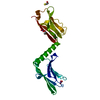
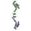
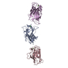
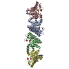
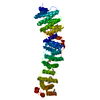
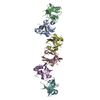
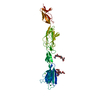
CRYSTAL STRUCTURE OF THE MYOMESIN DOMAINS MY9-MY11
Components
MYOMESIN
Keywords
STRUCTURAL PROTEIN / SARCOMERE / M-BAND / IMMUNOGLOBULIN- LIKE DOMAIN
Function / homology
Function and homology information
extraocular skeletal muscle development / striated muscle myosin thick filament / : / M band / sarcomere organization / structural constituent of muscle / positive regulation of protein secretion / kinase binding / positive regulation of gene expression / protein homodimerization activity / identical protein binding Similarity search - Function
: / Immunoglobulin I-set / Immunoglobulin I-set domain / Fibronectin type III domain / Fibronectin type 3 domain / Immunoglobulin subtype 2 / Immunoglobulin C-2 Type / Fibronectin type-III domain profile. / Fibronectin type III / Fibronectin type III superfamily ...: / Immunoglobulin I-set / Immunoglobulin I-set domain / Fibronectin type III domain / Fibronectin type 3 domain / Immunoglobulin subtype 2 / Immunoglobulin C-2 Type / Fibronectin type-III domain profile. / Fibronectin type III / Fibronectin type III superfamily / Immunoglobulin subtype / Immunoglobulin / Ig-like domain profile. / Immunoglobulin-like domain / Immunoglobulin-like domain superfamily / Immunoglobulin-like fold / Immunoglobulins / Immunoglobulin-like / Sandwich / Mainly Beta Similarity search - Domain/homology
Resolution: 2.5→25 Å / Cor.coef. Fo:Fc: 0.948 / Cor.coef. Fo:Fc free: 0.928 / SU B: 23.84 / SU ML: 0.283 / Cross valid method: THROUGHOUT / ESU R: 0.434 / ESU R Free: 0.292 / Stereochemistry target values: MAXIMUM LIKELIHOOD / Details: HYDROGENS HAVE BEEN ADDED IN THE RIDING POSITIONS.
Rfactor
Num. reflection
% reflection
Selection details
Rfree
0.271
465
3.1 %
RANDOM
Rwork
0.20934
-
-
-
obs
0.21127
14571
100 %
-
Solvent computation
Ion probe radii: 0.8 Å / Shrinkage radii: 0.8 Å / VDW probe radii: 1.2 Å / Solvent model: BABINET MODEL WITH MASK
 Movie
Movie Controller


 Open data
 Basic information
 Components
 Keywords
 Function and homology information
 HOMO SAPIENS (human)
 X-RAY DIFFRACTION /
  Authors
 Citation
 Structure visualization
 Downloads & links
 Other downloads
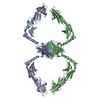


 PDBj
PDBj


 Assembly


 Mass: 78.133 Da / Num. of mol.: 3 / Source method: obtained synthetically / Formula: C2H6OS
 Mass: 120.147 Da / Num. of mol.: 1 / Source method: obtained synthetically / Formula: C5H12O3 / Comment: inhibitor, precipitant*YM
 Mass: 18.015 Da / Num. of mol.: 182 / Source method: isolated from a natural source / Formula: H2O
 Sample preparation
 / Beamline: X12 / Wavelength: 0.97699
  Processing