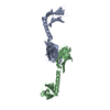
- PDB-2r15: Crystal structure of the myomesin domains 12 and 13 -
+
Open data
ID or keywords:
Loading...
-
Basic information
Entry
Database: PDB / ID: 2r15
Title
Crystal structure of the myomesin domains 12 and 13
Components
Myomesin-1
Keywords
CONTRACTILE PROTEIN / SARCOMERIC PROTEIN / IG-LIKE DOMAINS / HOMODIMER / Immunoglobulin domain / Muscle protein / Thick filament
Function / homology
Function and homology information
extraocular skeletal muscle development / striated muscle myosin thick filament / : / M band / sarcomere organization / structural constituent of muscle / positive regulation of protein secretion / kinase binding / positive regulation of gene expression / protein homodimerization activity / identical protein binding Similarity search - Function
: / Immunoglobulin I-set / Immunoglobulin I-set domain / Fibronectin type III domain / Fibronectin type 3 domain / Immunoglobulin subtype 2 / Immunoglobulin C-2 Type / Fibronectin type-III domain profile. / Fibronectin type III / Fibronectin type III superfamily ...: / Immunoglobulin I-set / Immunoglobulin I-set domain / Fibronectin type III domain / Fibronectin type 3 domain / Immunoglobulin subtype 2 / Immunoglobulin C-2 Type / Fibronectin type-III domain profile. / Fibronectin type III / Fibronectin type III superfamily / Immunoglobulin subtype / Immunoglobulin / Ig-like domain profile. / Immunoglobulin-like domain / Immunoglobulin-like domain superfamily / Immunoglobulin-like fold / Immunoglobulins / Immunoglobulin-like / Sandwich / Mainly Beta Similarity search - Domain/homology
Journal: EMBO J / Year: 2008 Title: Molecular basis of the C-terminal tail-to-tail assembly of the sarcomeric filament protein myomesin. Authors: Nikos Pinotsis / Stephan Lange / Jean-Claude Perriard / Dmitri I Svergun / Matthias Wilmanns / Abstract: Sarcomeric filament proteins display extraordinary properties in terms of protein length and mechanical elasticity, requiring specific anchoring and assembly mechanisms. To establish the molecular ...Sarcomeric filament proteins display extraordinary properties in terms of protein length and mechanical elasticity, requiring specific anchoring and assembly mechanisms. To establish the molecular basis of terminal filament assembly, we have selected the sarcomeric M-band protein myomesin as a prototypic filament model. The crystal structure of the myomesin C-terminus, comprising a tandem array of two immunoglobulin (Ig) domains My12 and My13, reveals a dimeric end-to-end filament of 14.3 nm length. Although the two domains share the same fold, an unexpected rearrangement of one beta-strand reveals how they are evolved into unrelated functions, terminal filament assembly (My13) and filament propagation (My12). The two domains are connected by a six-turn alpha-helix, of which two turns are void of any interactions with other protein parts. Thus, the overall structure of the assembled myomesin C-terminus resembles a three-body beads-on-the-string model with potentially elastic properties. We predict that the found My12-helix-My13 domain topology may provide a structural template for the filament architecture of the entire C-terminal Ig domain array My9-My13 of myomesin.
Mass: 18.015 Da / Num. of mol.: 247 / Source method: isolated from a natural source / Formula: H2O
-
Experimental details
-
Experiment
Experiment
Method: X-RAY DIFFRACTION / Number of used crystals: 2
-
Sample preparation
Crystal
Density Matthews: 2.96 Å3/Da / Density % sol: 58.48 %
Crystal grow
Temperature: 292 K / Method: vapor diffusion, hanging drop / pH: 7.5 Details: 0.16M CH3COONH4, 14% (W/V) PEG 20000, pH 7.50, VAPOR DIFFUSION, HANGING DROP, temperature 292K
-
Data collection
Diffraction
ID
Mean temperature (K)
Crystal-ID
1
100
1
2
100
1
Diffraction source
Source
Site
Beamline
ID
Wavelength (Å)
SYNCHROTRON
EMBL/DESY, HAMBURG
X11
1
0.8126, 0.91837, 0.8126
SYNCHROTRON
EMBL/DESY, HAMBURG
BW7A
2
0.98214, 0.98264, 0.91837
Detector
Type: MAR CCD 165 mm / Detector: CCD / Date: May 14, 2004
Radiation
Monochromator: GE SINGLE CRYSTAL CRYSTAL / Protocol: MAD / Monochromatic (M) / Laue (L): M / Scattering type: x-ray
Radiation wavelength
ID
Wavelength (Å)
Relative weight
1
0.8126
1
2
0.91837
1
3
0.98214
1
4
0.98264
1
Reflection
Resolution: 2.24→20 Å / Num. obs: 25757 / % possible obs: 95.7 % / Observed criterion σ(I): 0 / Redundancy: 4.9 % / Rmerge(I) obs: 0.076 / Net I/σ(I): 9.9
Reflection shell
Resolution: 2.24→2.28 Å / Redundancy: 4.4 % / Rmerge(I) obs: 0.382 / % possible all: 93.2
-
Processing
Software
Name
Version
Classification
CNS
refinement
REFMAC
5.2.0005
refinement
MAR345
datacollection
DENZO
datareduction
SCALEPACK
datascaling
CNS
phasing
Refinement
Method to determine structure: MAD / Resolution: 2.24→19.87 Å / Cor.coef. Fo:Fc: 0.955 / Cor.coef. Fo:Fc free: 0.942 / SU B: 13.663 / SU ML: 0.167 / TLS residual ADP flag: LIKELY RESIDUAL / Cross valid method: THROUGHOUT / σ(F): 0 / ESU R: 0.249 / ESU R Free: 0.188 / Stereochemistry target values: MAXIMUM LIKELIHOOD Details: THE MAD DATA SET WAS USED ONLY UP TO INITIAL DENSITY MODIFICATION
Rfactor
Num. reflection
% reflection
Selection details
Rfree
0.223
683
2.7 %
RANDOM
Rwork
0.196
-
-
-
obs
-
25697
100 %
-
Solvent computation
Ion probe radii: 0.8 Å / Shrinkage radii: 0.8 Å / VDW probe radii: 1.2 Å / Solvent model: BABINET MODEL WITH MASK
Displacement parameters
Biso mean: 44.68 Å2
Baniso -1
Baniso -2
Baniso -3
1-
-1.58 Å2
0 Å2
0 Å2
2-
-
2.09 Å2
0 Å2
3-
-
-
-0.51 Å2
Refinement step
Cycle: LAST / Resolution: 2.24→19.87 Å
Protein
Nucleic acid
Ligand
Solvent
Total
Num. atoms
3209
0
22
247
3478
Refine LS restraints
Refine-ID
Type
Dev ideal
Dev ideal target
Number
X-RAY DIFFRACTION
r_bond_refined_d
0.012
0.022
3304
X-RAY DIFFRACTION
r_angle_refined_deg
1.38
1.951
4480
X-RAY DIFFRACTION
r_dihedral_angle_1_deg
6.401
5
414
X-RAY DIFFRACTION
r_dihedral_angle_2_deg
36.155
25.4
150
X-RAY DIFFRACTION
r_dihedral_angle_3_deg
16.339
15
551
X-RAY DIFFRACTION
r_dihedral_angle_4_deg
22.087
15
12
X-RAY DIFFRACTION
r_chiral_restr
0.096
0.2
497
X-RAY DIFFRACTION
r_gen_planes_refined
0.005
0.02
2506
X-RAY DIFFRACTION
r_nbd_refined
0.219
0.2
1325
X-RAY DIFFRACTION
r_nbtor_refined
0.305
0.2
2196
X-RAY DIFFRACTION
r_xyhbond_nbd_refined
0.161
0.2
235
X-RAY DIFFRACTION
r_symmetry_vdw_refined
0.188
0.2
64
X-RAY DIFFRACTION
r_symmetry_hbond_refined
0.263
0.2
18
X-RAY DIFFRACTION
r_mcbond_it
1.506
3
2107
X-RAY DIFFRACTION
r_mcangle_it
2.442
5
3310
X-RAY DIFFRACTION
r_scbond_it
4.099
7
1368
X-RAY DIFFRACTION
r_scangle_it
6.192
10
1170
LS refinement shell
Resolution: 2.24→2.32 Å / Total num. of bins used: 15
Rfactor
Num. reflection
% reflection
Rfree
0.389
81
-
Rwork
0.363
2266
-
obs
-
-
100 %
Refinement TLS params.
Method: refined / Refine-ID: X-RAY DIFFRACTION
ID
L11 (°2)
L12 (°2)
L13 (°2)
L22 (°2)
L23 (°2)
L33 (°2)
S11 (Å °)
S12 (Å °)
S13 (Å °)
S21 (Å °)
S22 (Å °)
S23 (Å °)
S31 (Å °)
S32 (Å °)
S33 (Å °)
T11 (Å2)
T12 (Å2)
T13 (Å2)
T22 (Å2)
T23 (Å2)
T33 (Å2)
Origin x (Å)
Origin y (Å)
Origin z (Å)
1
9.9635
-2.0485
0.0964
9.3604
1.1613
15.1981
-0.4478
0.8098
-1.3819
0.4437
-0.0408
0.3755
1.3379
-0.2348
0.4886
0.2899
-0.0696
0.0623
-0.1032
-0.0513
-0.053
9.98
6.3251
90.8505
2
5.9755
-4.0087
-0.7989
10.2276
0.9271
6.2625
0.1803
0.6242
0.0418
-0.0103
-0.4809
-0.0654
0.0988
-0.3366
0.3006
0.0007
-0.0122
0.0196
-0.1001
-0.0452
-0.2436
7.0087
16.2704
90.5485
3
6.1538
-5.112
1.1025
11.8025
-1.345
5.2896
0.2493
0.2428
-0.9496
0.5572
-0.3456
0.1041
1.5491
0.17
0.0963
0.5136
0.057
0.0728
-0.0736
0.0547
0.001
11.2182
3.5417
98.1488
4
0.0578
0.7435
-1.8582
9.6467
-24.1215
60.3176
0.2281
0.2922
0.3723
0.4928
-0.0214
0.2176
-0.3408
0.4795
-0.2067
0.0039
0.0864
0.0481
-0.0335
0.0495
-0.091
5.3467
28.4052
87.1571
5
6.9657
2.1815
-2.2217
2.7764
-2.196
4.0286
-0.0512
-0.223
0.0808
-0.0663
0.0379
-0.0395
0.1593
-0.0114
0.0133
-0.3028
0.0383
-0.0138
-0.2843
0.0009
-0.2413
16.4236
42.2109
55.0016
6
3.8688
4.8926
-3.4228
9.143
-5.5895
3.5662
0.1914
-0.6509
1.194
0.5483
0.474
0.4772
-0.7455
-0.5853
-0.6654
-0.0629
0.1206
0.0254
-0.0975
-0.0332
0.06
13.145
51.7934
61.1166
7
8.4497
2.5722
-4.0154
3.4298
-2.0799
3.6399
-0.0453
-0.0609
0.3416
0.1811
0.1417
-0.0733
-0.2495
-0.2834
-0.0963
-0.2781
0.0429
-0.0289
-0.3225
-0.0303
-0.1552
16.383
47.5072
57.275
8
12.7119
-1.3236
-2.0196
3.1295
1.7419
9.9029
-0.0541
-1.5536
1.7595
0.2729
0.1989
0.0623
-1.3796
0.9017
-0.1448
0.4449
-0.0493
-0.037
0.0455
-0.1051
0.1301
10.8012
83.1218
17.1042
9
1.6314
0.5849
2.3603
6.667
2.2422
8.6085
0.0107
-0.152
0.0817
0.14
0.2053
-0.3228
-0.1897
0.1908
-0.216
0.1243
-0.0361
-0.0256
-0.0453
-0.0014
-0.1146
7.3476
73.4086
15.0337
10
18.6503
15.5535
7.2143
21.5752
1.9635
11.3935
-0.4784
0.0426
2.0162
-0.4926
-0.0989
0.2813
-2.3811
1.3573
0.5773
0.5895
-0.2098
-0.0357
0.0741
0.0056
0.3292
11.456
87.7241
10.8252
11
8.1745
-12.201
13.9238
24.1312
-29.3601
36.1449
-0.1306
-0.5899
-0.166
-0.6679
0.1383
0.4883
0.3189
0.4762
-0.0077
0.1406
0.0137
-0.1465
-0.098
0.0344
-0.1222
5.8928
60.7911
14.3573
12
5.5815
-1.1858
0.9787
3.0344
-1.0109
3.0966
-0.2386
0.4472
0.3084
0.0762
-0.0272
-0.2483
-0.1972
-0.2235
0.2658
-0.3307
-0.0264
0.0107
-0.2409
0.0069
-0.2079
17.2041
42.0098
44.2954
13
2.2404
-2.3827
2.001
6.8824
-4.1326
8.1626
0.0492
0.8823
-0.8139
-0.5446
0.3339
0.0691
0.6562
-0.2899
-0.3831
-0.1497
-0.1018
-0.016
-0.0203
-0.0594
-0.0169
15.1631
32.4403
37.4675
14
8.7926
-1.8623
3.2189
3.0477
-0.896
2.5782
-0.0813
0.6037
-0.2746
-0.2114
0.1339
-0.1459
0.0469
-0.2696
-0.0526
-0.2924
-0.0471
0.0042
-0.1967
-0.0497
-0.1944
17.6286
36.9333
41.92
Refinement TLS group
ID
Refine-ID
Refine TLS-ID
Auth asym-ID
Label asym-ID
Auth seq-ID
Label seq-ID
1
X-RAY DIFFRACTION
1
A
A
1461 - 1466
6 - 11
2
X-RAY DIFFRACTION
1
A
A
1479 - 1510
24 - 55
3
X-RAY DIFFRACTION
2
A
A
1467 - 1478
12 - 23
4
X-RAY DIFFRACTION
2
A
A
1511 - 1528
56 - 73
5
X-RAY DIFFRACTION
3
A
A
1529 - 1546
74 - 91
6
X-RAY DIFFRACTION
4
A
A
1547 - 1571
92 - 116
7
X-RAY DIFFRACTION
5
A
A
1572 - 1599
117 - 144
8
X-RAY DIFFRACTION
6
A
A
1600 - 1620
145 - 165
9
X-RAY DIFFRACTION
7
A
A
1621 - 1667
166 - 212
10
X-RAY DIFFRACTION
8
B
B
1461 - 1466
6 - 11
11
X-RAY DIFFRACTION
8
B
B
1479 - 1510
24 - 55
12
X-RAY DIFFRACTION
9
B
B
1467 - 1478
12 - 23
13
X-RAY DIFFRACTION
9
B
B
1511 - 1528
56 - 73
14
X-RAY DIFFRACTION
10
B
B
1529 - 1546
74 - 91
15
X-RAY DIFFRACTION
11
B
B
1547 - 1571
92 - 116
16
X-RAY DIFFRACTION
12
B
B
1572 - 1599
117 - 144
17
X-RAY DIFFRACTION
13
B
B
1600 - 1620
145 - 165
18
X-RAY DIFFRACTION
14
B
B
1621 - 1667
166 - 212
+
About Yorodumi
-
News
-
Feb 9, 2022. New format data for meta-information of EMDB entries
New format data for meta-information of EMDB entries
Version 3 of the EMDB header file is now the official format.
The previous official version 1.9 will be removed from the archive.
In the structure databanks used in Yorodumi, some data are registered as the other names, "COVID-19 virus" and "2019-nCoV". Here are the details of the virus and the list of structure data.
Jan 31, 2019. EMDB accession codes are about to change! (news from PDBe EMDB page)
EMDB accession codes are about to change! (news from PDBe EMDB page)
The allocation of 4 digits for EMDB accession codes will soon come to an end. Whilst these codes will remain in use, new EMDB accession codes will include an additional digit and will expand incrementally as the available range of codes is exhausted. The current 4-digit format prefixed with “EMD-” (i.e. EMD-XXXX) will advance to a 5-digit format (i.e. EMD-XXXXX), and so on. It is currently estimated that the 4-digit codes will be depleted around Spring 2019, at which point the 5-digit format will come into force.
The EM Navigator/Yorodumi systems omit the EMD- prefix.
Related info.:Q: What is EMD? / ID/Accession-code notation in Yorodumi/EM Navigator
Yorodumi is a browser for structure data from EMDB, PDB, SASBDB, etc.
This page is also the successor to EM Navigator detail page, and also detail information page/front-end page for Omokage search.
The word "yorodu" (or yorozu) is an old Japanese word meaning "ten thousand". "mi" (miru) is to see.
Related info.:EMDB / PDB / SASBDB / Comparison of 3 databanks / Yorodumi Search / Aug 31, 2016. New EM Navigator & Yorodumi / Yorodumi Papers / Jmol/JSmol / Function and homology information / Changes in new EM Navigator and Yorodumi
 Movie
Movie Controller


 Open data
 Basic information
 Components
 Keywords
 Function and homology information
 Homo sapiens (human)
 X-RAY DIFFRACTION /
  Authors
 Citation
 Structure visualization
 Downloads & links
 Other downloads


 PDBj
PDBj


 Assembly


 Mass: 59.044 Da / Num. of mol.: 1 / Source method: obtained synthetically / Formula: C2H3O2
 Mass: 92.094 Da / Num. of mol.: 3 / Source method: obtained synthetically / Formula: C3H8O3
 Mass: 18.015 Da / Num. of mol.: 247 / Source method: isolated from a natural source / Formula: H2O
 Sample preparation
 Processing