 Movie
Movie Controller

[English] 日本語
 Yorodumi
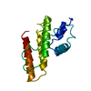
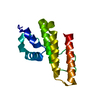
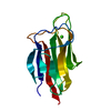
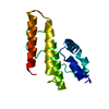
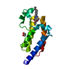
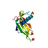
- PDB-1lwb: Crystal structure of prokaryotic phospholipase A2 at atomic resolution -
+ Open data

- Basic information
| Entry | Database: PDB / ID: 1lwb | ||||||
|---|---|---|---|---|---|---|---|
| Title | Crystal structure of prokaryotic phospholipase A2 at atomic resolution | ||||||
 Components Components | putative secreted protein | ||||||
 Keywords Keywords | HYDROLASE / Phospholipase A2 / atomic resolution / internal motion | ||||||
| Function / homology |  Function and homology information Function and homology informationA2-type glycerophospholipase activity / arachidonate secretion / phospholipid metabolic process / metal ion binding Similarity search - Function | ||||||
| Biological species |  Streptomyces violaceoruber (bacteria) Streptomyces violaceoruber (bacteria) | ||||||
| Method |  X-RAY DIFFRACTION / MOLECULAR REPLACEMENT / Resolution: 1.05 Å X-RAY DIFFRACTION / MOLECULAR REPLACEMENT / Resolution: 1.05 Å | ||||||
 Authors Authors | Matoba, Y. / Sugiyama, M. | ||||||
 Citation Citation | Journal: PROTEINS: STRUCT.,FUNCT.,GENET. / Year: 2003 Title: Atomic resolution structure of prokaryotic phospholipase A2: Analysis of internal motion and implication for a catalytic mechanism. Authors: Matoba, Y. / Sugiyama, M. #1: Journal: J.Biol.Chem. / Year: 2002Title: The crystal structure of prokaryotic phospholipase A2 Authors: Matoba, Y. / Katsube, Y. / Sugiyama, M. #2: Journal: J.Biol.Chem. / Year: 2002Title: A novel prokaryotic phospholipase A2. Characterizaton, gene cloning and solution structure Authors: Sugiyama, M. / Ohtani, K. / Izuhara, M. / Koike, T. / Suzuki, K. / Imamura, S. / Misaki, H. | ||||||
| History |
|
- Structure visualization
| Structure viewer | Molecule: MolmilJmol/JSmol |
|---|
- Downloads & links
-Download
| PDBx/mmCIF format | 1lwb.cif.gz | 87.8 KB | Display | PDBx/mmCIF format |
|---|---|---|---|---|
| PDB format | pdb1lwb.ent.gz | 67.8 KB | Display | PDB format |
| PDBx/mmJSON format | 1lwb.json.gz | Tree view | PDBx/mmJSON format | |
| Others |  Other downloads Other downloads |
-Validation report
| Arichive directory | https://data.pdbj.org/pub/pdb/validation_reports/lw/1lwbftp://data.pdbj.org/pub/pdb/validation_reports/lw/1lwb | HTTPS FTP |
|---|
-Related structure data
| Related structure data |  1fazS S: Starting model for refinement |
|---|---|
| Similar structure data |


-Links
 PDBj
PDBj
- Assembly
| Deposited unit | 
| ||||||||
|---|---|---|---|---|---|---|---|---|---|
| 1 |
| ||||||||
| Unit cell |
| ||||||||
| Details | This enzyme is a monomeric protein in solution. |
-Components
| #1: Protein | Mass: 13570.782 Da / Num. of mol.: 1 Source method: isolated from a genetically manipulated source Source: (gene. exp.) Streptomyces violaceoruber (bacteria) / Plasmid: pIJ680 / Species (production host): Streptomyces lividans / Production host: Streptomyces lividans TK24 (bacteria) / Strain (production host): TK24References: UniProt: Q9Z4W2, UniProt: Q6UV28*PLUS, phospholipase A2 |
|---|---|
| #2: Water | ChemComp-HOH /  Mass: 18.015 Da / Num. of mol.: 177 / Source method: isolated from a natural source / Formula: H2O Mass: 18.015 Da / Num. of mol.: 177 / Source method: isolated from a natural source / Formula: H2O |
| Has protein modification | Y |
-Experimental details
-Experiment
| Experiment | Method: X-RAY DIFFRACTION / Number of used crystals: 1 |
|---|
- Sample preparation
| Crystal | Density Matthews: 1.48 Å3/Da / Density % sol: 17.1 % | ||||||||||||||||||||||||||||||||||||||||||
|---|---|---|---|---|---|---|---|---|---|---|---|---|---|---|---|---|---|---|---|---|---|---|---|---|---|---|---|---|---|---|---|---|---|---|---|---|---|---|---|---|---|---|---|
| Crystal grow | Temperature: 297 K / Method: vapor diffusion, hanging drop / pH: 6 Details: PEG 6000, lithium sulfate, sodium cacodylate, VAPOR DIFFUSION, HANGING DROP, temperature 297K | ||||||||||||||||||||||||||||||||||||||||||
| Crystal grow | *PLUS Temperature: 297 K / pH: 8 / Method: vapor diffusion, hanging drop / Details: Matoba, Y., (2002) J.Biol.Chem., 277, 20059. | ||||||||||||||||||||||||||||||||||||||||||
| Components of the solutions | *PLUS
|
-Data collection
| Diffraction | Mean temperature: 297 K |
|---|---|
| Diffraction source | Source: ROTATING ANODE / Type: RIGAKU RU200 / Wavelength: 0.71073 Å |
| Detector | Type: RIGAKU RAXIS IV / Detector: IMAGE PLATE / Date: 1997 |
| Radiation | Monochromator: GRAPHITE / Protocol: SINGLE WAVELENGTH / Monochromatic (M) / Laue (L): M / Scattering type: x-ray |
| Radiation wavelength | Wavelength: 0.71073 Å / Relative weight: 1 |
| Reflection | Resolution: 0.923→100 Å / Num. obs: 45532 / % possible obs: 67.48 % / Observed criterion σ(F): 2 / Observed criterion σ(I): 1 / Redundancy: 2.65 % / Rmerge(I) obs: 0.072 / Net I/σ(I): 8.21 |
| Reflection shell | Resolution: 0.923→0.954 Å / Rmerge(I) obs: 0.228 / Mean I/σ(I) obs: 1.64 / Num. unique all: 1418 / % possible all: 22.75 |
| Reflection shell | *PLUS % possible obs: 22.75 % / Num. unique obs: 1418 / Mean I/σ(I) obs: 5.82 |
- Processing
| Software |
| |||||||||||||||||||||||||||||||||
|---|---|---|---|---|---|---|---|---|---|---|---|---|---|---|---|---|---|---|---|---|---|---|---|---|---|---|---|---|---|---|---|---|---|---|
| Refinement | Method to determine structure: MOLECULAR REPLACEMENT Starting model: PDB ENTRY 1FAZ Resolution: 1.05→10 Å / Num. parameters: 10812 / Num. restraintsaints: 13414 / Cross valid method: FREE R / σ(F): 0 / Stereochemistry target values: Engh & Huber / Details: ANISOTROPIC TEMPERATURE FACTOR WAS INTRODUCED.
| |||||||||||||||||||||||||||||||||
| Refine analyze | Num. disordered residues: 34 / Occupancy sum hydrogen: 844 / Occupancy sum non hydrogen: 1120.7 | |||||||||||||||||||||||||||||||||
| Refinement step | Cycle: LAST / Resolution: 1.05→10 Å
| |||||||||||||||||||||||||||||||||
| Refine LS restraints |
| |||||||||||||||||||||||||||||||||
| Software | *PLUS Name: SHELXL / Version: 97 / Classification: refinement | |||||||||||||||||||||||||||||||||
| Refinement | *PLUS Rfactor all: 0.1037 / Rfactor obs: 0.103 / Rfactor Rwork: 0.097 | |||||||||||||||||||||||||||||||||
| Solvent computation | *PLUS | |||||||||||||||||||||||||||||||||
| Displacement parameters | *PLUS | |||||||||||||||||||||||||||||||||
| Refine LS restraints | *PLUS
|
