 Movie
Movie Controller

[English] 日本語
 Yorodumi
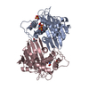
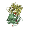
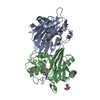
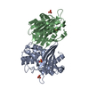
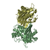
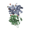
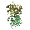
- PDB-1k4f: CRYSTAL STRUCTURE OF THE CLASS D BETA-LACTAMASE OXA-10 AT 1.6 A R... -
+ Open data

- Basic information
| Entry | Database: PDB / ID: 1k4f | ||||||
|---|---|---|---|---|---|---|---|
| Title | CRYSTAL STRUCTURE OF THE CLASS D BETA-LACTAMASE OXA-10 AT 1.6 A RESOLUTION | ||||||
 Components Components | Beta-lactamase PSE-2 | ||||||
 Keywords Keywords |  HYDROLASE / beta-lactamase / class D / carbamylated lysine HYDROLASE / beta-lactamase / class D / carbamylated lysine | ||||||
| Function / homology |  Function and homology informationpenicillin binding / antibiotic catabolic process / beta-lactamase activity / beta-lactamase / response to antibiotic Function and homology informationpenicillin binding / antibiotic catabolic process / beta-lactamase activity / beta-lactamase / response to antibioticSimilarity search - Function | ||||||
| Biological species |   Pseudomonas aeruginosa (bacteria) Pseudomonas aeruginosa (bacteria) | ||||||
| Method | X-RAY DIFFRACTION / SYNCHROTRON / MOLECULAR REPLACEMENT / Resolution: 1.6 Å | ||||||
 Authors Authors | Kerff, F. / Fonze, E. / Bouillene, F. / Frere, J.M. / Charlier, P. | ||||||
 Citation Citation | Journal: To be Published Title: Structure of the class D beta-lactamase OXA-2 Authors: Kerff, F. / Fonze, E. / Bouillene, F. / Frere, J.M. / Charlier, P. | ||||||
| History |
|
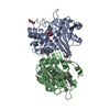
- Structure visualization
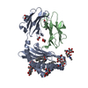
| Structure viewer | Molecule: MolmilJmol/JSmol |
|---|
- Downloads & links
-Download
| PDBx/mmCIF format | 1k4f.cif.gz | 127 KB | Display | PDBx/mmCIF format |
|---|---|---|---|---|
| PDB format | pdb1k4f.ent.gz | 97.5 KB | Display | PDB format |
| PDBx/mmJSON format | 1k4f.json.gz | Tree view | PDBx/mmJSON format | |
| Others |  Other downloads Other downloads |
-Validation report
| Arichive directory | https://data.pdbj.org/pub/pdb/validation_reports/k4/1k4fftp://data.pdbj.org/pub/pdb/validation_reports/k4/1k4f | HTTPS FTP |
|---|
-Related structure data
| Related structure data |  1k4eS S: Starting model for refinement |
|---|---|
| Similar structure data |


-Links
 PDBj
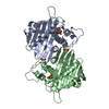
PDBj- Assembly
| Deposited unit | 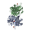
| ||||||||
|---|---|---|---|---|---|---|---|---|---|
| 1 |
| ||||||||
| Unit cell |
| ||||||||
| Details | The biological assembly is a dimer in the asymmetric unit formed by chains A and B |
-Components
| #1: Protein | Mass: 27755.541 Da / Num. of mol.: 2 Source method: isolated from a genetically manipulated source Source: (gene. exp.) Pseudomonas aeruginosa (bacteria) / Gene: plasmid pMON234 / Plasmid: pET22b / Species (production host): Escherichia coli / Production host: Escherichia coli BL21(DE3) (bacteria) / Strain (production host): BL21(DE3) / References: UniProt: P14489, beta-lactamase#2: Chemical | ChemComp-SO4 / Sulfate  Mass: 96.063 Da / Num. of mol.: 4 / Source method: obtained synthetically / Formula: SO4 Mass: 96.063 Da / Num. of mol.: 4 / Source method: obtained synthetically / Formula: SO4#3: Water | ChemComp-HOH / | Water Mass: 18.015 Da / Num. of mol.: 626 / Source method: isolated from a natural source / Formula: H2O Mass: 18.015 Da / Num. of mol.: 626 / Source method: isolated from a natural source / Formula: H2O |
|---|
-Experimental details
-Experiment
| Experiment | Method: X-RAY DIFFRACTION / Number of used crystals: 1 |
|---|
- Sample preparation
| Crystal | Density Matthews: 2.59 Å3/Da / Density % sol: 52.49 % |
|---|---|
| Crystal grow | Temperature: 293 K / Method: vapor diffusion, hanging drop / pH: 9 Details: ammonium sulfate, bicine, pH 9, VAPOR DIFFUSION, HANGING DROP, temperature 293K |
-Data collection
| Diffraction | Mean temperature: 100 K |
|---|---|
| Diffraction source | Source: SYNCHROTRON / Site: ESRF  / Beamline: ID14-1 / Wavelength: 0.934 Å / Beamline: ID14-1 / Wavelength: 0.934 Å |
| Detector | Type: MARRESEARCH / Detector: CCD / Date: Jun 7, 2000 |
| Radiation | Monochromator: Diamond 111 / Protocol: SINGLE WAVELENGTH / Monochromatic (M) / Laue (L): M / Scattering type: x-ray |
| Radiation wavelength | Wavelength: 0.934 Å / Relative weight: 1 |
| Reflection | Resolution: 1.6→32.6 Å / Num. all: 74910 / Num. obs: 74815 / % possible obs: 97.7 % / Observed criterion σ(F): 0 / Observed criterion σ(I): 0 / Redundancy: 3.9 % / Biso Wilson estimate: 17 Å2 / Rsym value: 0.047 / Net I/σ(I): 10.3 |
| Reflection shell | Resolution: 1.6→1.69 Å / Redundancy: 3.4 % / Mean I/σ(I) obs: 7.2 / Num. unique all: 9515 / Rsym value: 0.093 / % possible all: 95.7 |
- Processing
| Software |
| ||||||||||||||||||||||||||||||||||||
|---|---|---|---|---|---|---|---|---|---|---|---|---|---|---|---|---|---|---|---|---|---|---|---|---|---|---|---|---|---|---|---|---|---|---|---|---|---|
| Refinement | Method to determine structure: MOLECULAR REPLACEMENT Starting model: PDB ENTRY 1K4E Resolution: 1.6→26.39 Å / Rfactor Rfree error: 0.003 / Data cutoff high absF: 1578368.11 / Data cutoff low absF: 0 / Isotropic thermal model: RESTRAINED / Cross valid method: THROUGHOUT / σ(F): 2 / Stereochemistry target values: Engh & Huber
| ||||||||||||||||||||||||||||||||||||
| Solvent computation | Solvent model: FLAT MODEL / Bsol: 69.7252 Å2 / ksol: 0.408073 e/Å3 | ||||||||||||||||||||||||||||||||||||
| Displacement parameters | Biso mean: 20.5 Å2
| ||||||||||||||||||||||||||||||||||||
| Refine analyze |
| ||||||||||||||||||||||||||||||||||||
| Refinement step | Cycle: LAST / Resolution: 1.6→26.39 Å
| ||||||||||||||||||||||||||||||||||||
| Refine LS restraints |
| ||||||||||||||||||||||||||||||||||||
| LS refinement shell | Resolution: 1.6→1.66 Å / Rfactor Rfree error: 0.011 / Total num. of bins used: 10
| ||||||||||||||||||||||||||||||||||||
| Xplor file |
|
