 Movie
Movie Controller


+ Open data

- Basic information
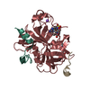
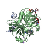
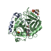
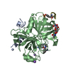
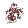
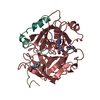
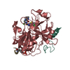
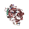
| Entry | Database: PDB / ID: 1k22 | |||||||||
|---|---|---|---|---|---|---|---|---|---|---|
| Title | HUMAN THROMBIN-INHIBITOR COMPLEX | |||||||||
 Components Components |
| |||||||||
 Keywords Keywords | HYDROLASE/HYDROLASE INHIBITOR / COMPLEX (SERINE PROTEASE-INHIBITOR) /  HYDROLASE / HYDROLASE-HYDROLASE INHIBITOR COMPLEX HYDROLASE / HYDROLASE-HYDROLASE INHIBITOR COMPLEX | |||||||||
| Function / homology |  Function and homology information Function and homology informationpositive regulation of lipid kinase activity / positive regulation of phospholipase C-activating G protein-coupled receptor signaling pathway / cytolysis by host of symbiont cells / thrombospondin receptor activity / Defective factor XII causes hereditary angioedema / thrombin / neutrophil-mediated killing of gram-negative bacterium / regulation of blood coagulation / ligand-gated ion channel signaling pathway / Defective F8 cleavage by thrombin ...positive regulation of lipid kinase activity / positive regulation of phospholipase C-activating G protein-coupled receptor signaling pathway / cytolysis by host of symbiont cells / thrombospondin receptor activity / Defective factor XII causes hereditary angioedema / thrombin / neutrophil-mediated killing of gram-negative bacterium / regulation of blood coagulation / ligand-gated ion channel signaling pathway / Defective F8 cleavage by thrombin / Platelet Aggregation (Plug Formation) / negative regulation of platelet activation / negative regulation of astrocyte differentiation / positive regulation of collagen biosynthetic process / negative regulation of cytokine production involved in inflammatory response / positive regulation of blood coagulation / negative regulation of fibrinolysis / Gamma-carboxylation of protein precursors / Transport of gamma-carboxylated protein precursors from the endoplasmic reticulum to the Golgi apparatus / Common Pathway of Fibrin Clot Formation / Removal of aminoterminal propeptides from gamma-carboxylated proteins / fibrinolysis / regulation of cytosolic calcium ion concentration / Intrinsic Pathway of Fibrin Clot Formation / Peptide ligand-binding receptors / positive regulation of release of sequestered calcium ion into cytosol / Regulation of Complement cascade / acute-phase response / Cell surface interactions at the vascular wall / lipopolysaccharide binding / negative regulation of proteolysis / positive regulation of receptor signaling pathway via JAK-STAT / growth factor activity / serine-type endopeptidase inhibitor activity / positive regulation of insulin secretion / platelet activation / response to wounding / Golgi lumen / positive regulation of protein localization to nucleus / Regulation of Insulin-like Growth Factor (IGF) transport and uptake by Insulin-like Growth Factor Binding Proteins (IGFBPs) / positive regulation of reactive oxygen species metabolic process / blood coagulation / antimicrobial humoral immune response mediated by antimicrobial peptide / Thrombin signalling through proteinase activated receptors (PARs) / heparin binding / regulation of cell shape / positive regulation of cell growth / G alpha (q) signalling events / collagen-containing extracellular matrix / blood microparticle / cell surface receptor signaling pathway / positive regulation of phosphatidylinositol 3-kinase/protein kinase B signal transduction / positive regulation of protein phosphorylation / G protein-coupled receptor signaling pathway / endoplasmic reticulum lumen / signaling receptor binding / serine-type endopeptidase activity / calcium ion binding / positive regulation of cell population proliferation / proteolysis / extracellular space / extracellular exosome / extracellular region / plasma membraneSimilarity search - Function | |||||||||
| Biological species |  Homo sapiens (human) Homo sapiens (human) Hirudo medicinalis (medicinal leech) Hirudo medicinalis (medicinal leech) | |||||||||
| Method | X-RAY DIFFRACTION / OTHER / Resolution: 1.93 Å | |||||||||
 Authors Authors | Stubbs, M.T. / Musil, D. | |||||||||
 Citation Citation | Journal: J.Mol.Biol. / Year: 2001 Title: Factorising ligand affinity: a combined thermodynamic and crystallographic study of trypsin and thrombin inhibition. Authors: Dullweber, F. / Stubbs, M.T. / Musil, D. / Sturzebecher, J. / Klebe, G. #1: Journal: J.Med.Chem. / Year: 1998Title: Structural and Functional Analyses of Benzamidine-Based Inhibitors in Complex with Trypsin: Implications for the Inhibition of Factor Xa, Tpa, and Urokinase Authors: Renatus, M. / Bode, W. / Huber, R. / Stuerzebecher, J. / Stubbs, M.T. #2: Journal: FEBS Lett. / Year: 1995Title: Crystal Structures of Factor Xa Specific Inhibitors in Complex with Trypsin: Structural Grounds for Inhibition of Factor Xa and Selectivity Against Thrombin Authors: Stubbs, M.T. / Huber, R. / Bode, W. #3: Journal: Thromb.Res. / Year: 1993Title: A Player of Many Parts: The Spotlight Falls on Thrombin'S Structure Authors: Stubbs, M.T. / Bode, W. | |||||||||
| History |
|
- Structure visualization
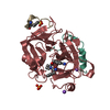
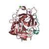
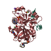
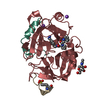
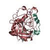
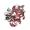
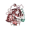
| Structure viewer | Molecule: MolmilJmol/JSmol |
|---|
- Downloads & links
-Download
| PDBx/mmCIF format | 1k22.cif.gz | 78.4 KB | Display | PDBx/mmCIF format |
|---|---|---|---|---|
| PDB format | pdb1k22.ent.gz | 59.6 KB | Display | PDB format |
| PDBx/mmJSON format | 1k22.json.gz | Tree view | PDBx/mmJSON format | |
| Others |  Other downloads Other downloads |
-Validation report
| Arichive directory | https://data.pdbj.org/pub/pdb/validation_reports/k2/1k22ftp://data.pdbj.org/pub/pdb/validation_reports/k2/1k22 | HTTPS FTP |
|---|
-Related structure data
| Related structure data | 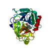 1k1iC 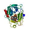 1k1jC 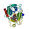 1k1lC 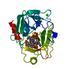 1k1mC 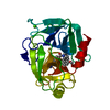 1k1nC  1k1oC 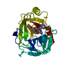 1k1pC 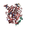 1k21C C: citing same article ( |
|---|---|
| Similar structure data |


-Links
 PDBj
PDBj


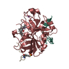
- Assembly
| Deposited unit | 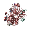
| ||||||||
|---|---|---|---|---|---|---|---|---|---|
| 1 |
| ||||||||
| Unit cell |
|
-Components
-Protein/peptide , 2 types, 2 molecules LI
| #1: Protein/peptide | Thrombin / ALPHA-THROMBIN / Coagulation factor II Mass: 4096.534 Da / Num. of mol.: 1 / Fragment: THROMBIN LIGHT CHAIN, Residues 323-363 / Source method: isolated from a natural source / Source: (natural) Homo sapiens (human) / References: UniProt: P00734, thrombin |
|---|---|
| #3: Protein/peptide | Mass: 1534.554 Da / Num. of mol.: 1 / Fragment: Residues 60-71 / Source method: isolated from a natural source / Source: (natural) Hirudo medicinalis (medicinal leech) / References: UniProt: P09945 |
-Protein / Sugars , 2 types, 2 molecules H
| #2: Protein | Thrombin / ALPHA-THROMBIN / Coagulation factor II Mass: 29780.219 Da / Num. of mol.: 1 / Fragment: THROMBIN HEAVY CHAIN, Residues 364-622 / Source method: isolated from a natural source / Source: (natural) Homo sapiens (human) / References: UniProt: P00734, thrombin |
|---|---|
| #4: Polysaccharide | 2-acetamido-2-deoxy-beta-D-glucopyranose-(1-4)-2-acetamido-2-deoxy-beta-D-glucopyranose / Mass: 424.401 Da / Num. of mol.: 1 Source method: isolated from a genetically manipulated source |
-Non-polymers , 3 types, 242 molecules 
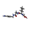

| #5: Chemical |  Mass: 22.990 Da / Num. of mol.: 2 / Source method: obtained synthetically / Formula: Na Mass: 22.990 Da / Num. of mol.: 2 / Source method: obtained synthetically / Formula: Na#6: Chemical | ChemComp-MEL / [(( |  Mass: 429.513 Da / Num. of mol.: 1 / Source method: obtained synthetically / Formula: C22H31N5O4 Mass: 429.513 Da / Num. of mol.: 1 / Source method: obtained synthetically / Formula: C22H31N5O4#7: Water | ChemComp-HOH / | WaterMass: 18.015 Da / Num. of mol.: 239 / Source method: isolated from a natural source / Formula: H2O |
|---|
-Details
| Compound details | CHYMOTRYPSIN NUMBERING (RATHER THAN SEQUENTIAL) SYSTEM IS USED, BASED ON THE TOPOLOGICAL ALIGNMENT ...CHYMOTRYPS | ||
|---|---|---|---|
| Nonpolymer details | HETATM MEL CORRESPOND| Sequence details | THROMBIN IS CLEAVED BETWEEN RESIDUES 15 AND 16. CHAIN IDENTIFIER *L* IS USED FOR RESIDUES 1H - 15 ...THROMBIN IS CLEAVED BETWEEN RESIDUES 15 AND 16. CHAIN IDENTIFIER | |
-Experimental details
-Experiment
| Experiment | Method: X-RAY DIFFRACTION / Number of used crystals: 1 |
|---|
- Sample preparation
| Crystal | Density Matthews: 2.48 Å3/Da / Density % sol: 50.47 % |
|---|
-Data collection
| Diffraction | Mean temperature: 287 K |
|---|---|
| Diffraction source | Source: ROTATING ANODE / Type: RIGAKU RU300 / Wavelength: 1.5418 |
| Detector | Type: RIGAKU RAXIS IV / Detector: IMAGE PLATE / Date: Jun 15, 1999 / Details: MIRRORS |
| Radiation | Monochromator: NI FILTER / Protocol: SINGLE WAVELENGTH / Monochromatic (M) / Laue (L): M / Scattering type: x-ray |
| Radiation wavelength | Wavelength: 1.5418 Å / Relative weight: 1 |
| Reflection | Resolution: 1.93→20 Å / Num. obs: 25984 / % possible obs: 99.8 % / Observed criterion σ(I): 0 / Redundancy: 3.9 % / Rmerge(I) obs: 0.054 / Rsym value: 0.054 |
| Reflection shell | Highest resolution: 1.93 Å / Rmerge(I) obs: 0.298 / Rsym value: 0.298 / % possible all: 98.3 |
- Processing
| Software |
| ||||||||||||||||||||||||||||||||||||||||||||||||||||||||||||
|---|---|---|---|---|---|---|---|---|---|---|---|---|---|---|---|---|---|---|---|---|---|---|---|---|---|---|---|---|---|---|---|---|---|---|---|---|---|---|---|---|---|---|---|---|---|---|---|---|---|---|---|---|---|---|---|---|---|---|---|---|---|
| Refinement | Method to determine structure: OTHER / Resolution: 1.93→500 Å / Data cutoff high absF: 10000000 / Data cutoff low absF: 0.001 / σ(F): 0
| ||||||||||||||||||||||||||||||||||||||||||||||||||||||||||||
| Refine analyze | Luzzati d res low obs: 10 Å | ||||||||||||||||||||||||||||||||||||||||||||||||||||||||||||
| Refinement step | Cycle: LAST / Resolution: 1.93→500 Å
| ||||||||||||||||||||||||||||||||||||||||||||||||||||||||||||
| Refine LS restraints |
| ||||||||||||||||||||||||||||||||||||||||||||||||||||||||||||
| LS refinement shell | Highest resolution: 1.93 Å | ||||||||||||||||||||||||||||||||||||||||||||||||||||||||||||
| Xplor file |
|
