 Movie
Movie Controller

[English] 日本語
 Yorodumi
- PDB-1jq7: HCMV protease dimer-interface mutant, S225Y complexed to Inhibito... -
+ Open data

- Basic information
| Entry | Database: PDB / ID: 1jq7 | ||||||
|---|---|---|---|---|---|---|---|
| Title | HCMV protease dimer-interface mutant, S225Y complexed to Inhibitor BILC 408 | ||||||
 Components Components | ASSEMBLIN | ||||||
 Keywords Keywords | HYDROLASE/HYDROLASE INHIBITOR / Herpesvirus / cytomegalovirus / serine protease / dimerization / enzyme activity regulation / HYDROLASE-HYDROLASE INHIBITOR COMPLEX | ||||||
| Function / homology |  Function and homology informationassemblin / viral release from host cell / host cell cytoplasm / serine-type endopeptidase activity / host cell nucleus / proteolysis / identical protein binding Function and homology informationassemblin / viral release from host cell / host cell cytoplasm / serine-type endopeptidase activity / host cell nucleus / proteolysis / identical protein bindingSimilarity search - Function | ||||||
| Biological species |   Human herpesvirus 5 Human herpesvirus 5 | ||||||
| Method | X-RAY DIFFRACTION / SYNCHROTRON / MOLECULAR REPLACEMENT / Resolution: 3 Å | ||||||
 Authors Authors | Batra, R. / Khayat, R. / Tong, L. | ||||||
 Citation Citation | Journal: Nat.Struct.Biol. / Year: 2001 Title: Molecular mechanism for dimerization to regulate the catalytic activity of human cytomegalovirus protease. Authors: Batra, R. / Khayat, R. / Tong, L. | ||||||
| History |
|
- Structure visualization
| Structure viewer | Molecule: MolmilJmol/JSmol |
|---|
- Downloads & links
-Download
| PDBx/mmCIF format | 1jq7.cif.gz | 99.3 KB | Display | PDBx/mmCIF format |
|---|---|---|---|---|
| PDB format | pdb1jq7.ent.gz | 74.8 KB | Display | PDB format |
| PDBx/mmJSON format | 1jq7.json.gz | Tree view | PDBx/mmJSON format | |
| Others |  Other downloads Other downloads |
-Validation report
| Arichive directory | https://data.pdbj.org/pub/pdb/validation_reports/jq/1jq7ftp://data.pdbj.org/pub/pdb/validation_reports/jq/1jq7 | HTTPS FTP |
|---|
-Related structure data
| Related structure data |  1jq6C  2wpoS S: Starting model for refinement C: citing same article ( |
|---|---|
| Similar structure data |


-Links
 PDBj
PDBj- Assembly
| Deposited unit | 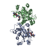
| ||||||||
|---|---|---|---|---|---|---|---|---|---|
| 1 |
| ||||||||
| Unit cell |
|
-Components
| #1: Protein | / PROTEASE Mass: 28206.641 Da / Num. of mol.: 2 / Mutation: A143Q, S225Y Source method: isolated from a genetically manipulated source Source: (gene. exp.) Human herpesvirus 5 / Production host:  Escherichia coli (E. coli) / References: UniProt: P16753, assemblin Escherichia coli (E. coli) / References: UniProt: P16753, assemblin#2: Chemical |   Type: peptide-like, Peptide-like / Class: Inhibitor / Mass: 717.939 Da / Num. of mol.: 2 / Source method: obtained synthetically / Formula: C37H63N7O7 / Details: This peptide was chemically synthesized. Type: peptide-like, Peptide-like / Class: Inhibitor / Mass: 717.939 Da / Num. of mol.: 2 / Source method: obtained synthetically / Formula: C37H63N7O7 / Details: This peptide was chemically synthesized.References: N-(6-aminohexanoyl)-3-methyl-L-valyl-3-methyl-L-valyl-N~4~,N~4~-dimethyl-N~1~-[(1R)-1-methyl-2,3-dioxo-3-{[(1S)-1- phenylpropyl]amino}propyl]-L-aspartamide Nonpolymer details | THE INHIBITOR 0FP IS COVALENTLY CONNECTED AT CARBON C4 TO THE ACTIVE SITE SERINES (A 1132 AND B ...THE INHIBITOR 0FP IS COVALENTLY | |
|---|
-Experimental details
-Experiment
| Experiment | Method: X-RAY DIFFRACTION / Number of used crystals: 1 |
|---|
- Sample preparation
| Crystal | Density Matthews: 2.58 Å3/Da / Density % sol: 52.4 % | |||||||||||||||||||||||||||||||||||||||||||||||||
|---|---|---|---|---|---|---|---|---|---|---|---|---|---|---|---|---|---|---|---|---|---|---|---|---|---|---|---|---|---|---|---|---|---|---|---|---|---|---|---|---|---|---|---|---|---|---|---|---|---|---|
| Crystal grow | Temperature: 294 K / Method: vapor diffusion, hanging drop / pH: 7.5 Details: PEG 4000, HEPES, sodium chloride, glycerol, spermine tetrahydrochloride, DTT, pH 7.5, VAPOR DIFFUSION, HANGING DROP, temperature 294K | |||||||||||||||||||||||||||||||||||||||||||||||||
| Crystal grow | *PLUS Temperature: 21 ℃ / Details: Tong, L., (1998) Nature Struct. Biol., 5, 819. | |||||||||||||||||||||||||||||||||||||||||||||||||
| Components of the solutions | *PLUS
|
-Data collection
| Diffraction | Mean temperature: 100 K |
|---|---|
| Diffraction source | Source: SYNCHROTRON / Site: NSLS  / Beamline: X8C / Wavelength: 0.978 Å / Beamline: X8C / Wavelength: 0.978 Å |
| Detector | Type: ADSC QUANTUM 4 / Detector: CCD / Date: Apr 2, 2001 |
| Radiation | Monochromator: Si 111 Channel / Protocol: SINGLE WAVELENGTH / Monochromatic (M) / Laue (L): M / Scattering type: x-ray |
| Radiation wavelength | Wavelength: 0.978 Å / Relative weight: 1 |
| Reflection | Resolution: 3→19.9 Å / Num. all: 48246 / Num. obs: 10900 / % possible obs: 85.5 % / Observed criterion σ(F): 7 / Observed criterion σ(I): 5 |
| Reflection shell | Resolution: 3→3.19 Å / % possible all: 97 |
| Reflection | *PLUS Num. measured all: 48246 / Rmerge(I) obs: 0.05 |
| Reflection shell | *PLUS Rmerge(I) obs: 0.129 |
- Processing
| Software |
| ||||||||||||||||||||||||||||||||||||||||
|---|---|---|---|---|---|---|---|---|---|---|---|---|---|---|---|---|---|---|---|---|---|---|---|---|---|---|---|---|---|---|---|---|---|---|---|---|---|---|---|---|---|
| Refinement | Method to determine structure: MOLECULAR REPLACEMENT Starting model: PDB ENTRY 2WPO (dimer) Resolution: 3→19.99 Å / Rfactor Rfree error: 0.012 / Data cutoff high absF: 2061042.99 / Data cutoff low absF: 0 / Isotropic thermal model: RESTRAINED / Cross valid method: THROUGHOUT / σ(F): 7 / Stereochemistry target values: Engh & Huber
| ||||||||||||||||||||||||||||||||||||||||
| Solvent computation | Solvent model: FLAT MODEL / Bsol: 10 Å2 / ksol: 0.250918 e/Å3 | ||||||||||||||||||||||||||||||||||||||||
| Displacement parameters | Biso mean: 28 Å2
| ||||||||||||||||||||||||||||||||||||||||
| Refine analyze |
| ||||||||||||||||||||||||||||||||||||||||
| Refinement step | Cycle: LAST / Resolution: 3→19.99 Å
| ||||||||||||||||||||||||||||||||||||||||
| Refine LS restraints |
| ||||||||||||||||||||||||||||||||||||||||
| LS refinement shell | Resolution: 3→3.19 Å / Rfactor Rfree error: 0.037 / Total num. of bins used: 6
| ||||||||||||||||||||||||||||||||||||||||
| Xplor file |
| ||||||||||||||||||||||||||||||||||||||||
| Software | *PLUS Name: CNS / Version: 1 / Classification: refinement | ||||||||||||||||||||||||||||||||||||||||
| Refinement | *PLUS σ(F): 7 / % reflection Rfree: 7.4 % / Rfactor obs: 0.26 / Rfactor Rwork: 0.26 | ||||||||||||||||||||||||||||||||||||||||
| Solvent computation | *PLUS | ||||||||||||||||||||||||||||||||||||||||
| Displacement parameters | *PLUS Biso mean: 28 Å2 | ||||||||||||||||||||||||||||||||||||||||
| Refine LS restraints | *PLUS
| ||||||||||||||||||||||||||||||||||||||||
| LS refinement shell | *PLUS Rfactor Rfree: 0.36 / % reflection Rfree: 7.5 % / Rfactor Rwork: 0.281 |
