 Movie
Movie Controller


+ Open data

- Basic information
| Entry | Database: PDB / ID: 2wpo | ||||||
|---|---|---|---|---|---|---|---|
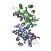
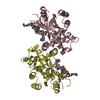
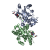
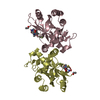
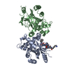
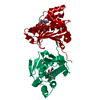
| Title | HCMV protease inhibitor complex | ||||||
 Components Components | HUMAN CYTOMEGALOVIRUS PROTEASE | ||||||
 Keywords Keywords | HYDROLASE/HYDROLASE INHIBITOR / VIRAL PROTEASE / HYDROLASE-HYDROLASE INHIBITOR complex / COAT PROTEIN / SERINE PROTEASE | ||||||
| Function / homology |  Function and homology information Function and homology informationassemblin / nuclear capsid assembly / viral release from host cell / host cell cytoplasm / serine-type endopeptidase activity / host cell nucleus / proteolysis / identical protein binding Similarity search - Function | ||||||
| Biological species |   Human herpesvirus 5 Human herpesvirus 5 | ||||||
| Method |  X-RAY DIFFRACTION / MOLECULAR REPLACEMENT / Resolution: 2.7 Å X-RAY DIFFRACTION / MOLECULAR REPLACEMENT / Resolution: 2.7 Å | ||||||
 Authors Authors | Tong, L. / Qian, C. / Massariol, M.-J. / Deziel, R. / Yoakim, C. / Lagace, L. | ||||||
 Citation Citation | Journal: Nat.Struct.Biol. / Year: 1998 Title: Conserved mode of peptidomimetic inhibition and substrate recognition of human cytomegalovirus protease. Authors: Tong, L. / Qian, C. / Massariol, M.J. / Deziel, R. / Yoakim, C. / Lagace, L. #1: Journal: Nature / Year: 1996Title: A New Serine-Protease Fold Revealed by the Crystal Structure of Human Cytomegalovirus Protease Authors: Tong, L. / Qian, C. / Massariol, M.J. / Bonneau, P.R. / Cordingley, M.G. / Lagace, L. | ||||||
| History |
|
- Structure visualization
| Structure viewer | Molecule: MolmilJmol/JSmol |
|---|
- Downloads & links
-Download
| PDBx/mmCIF format | 2wpo.cif.gz | 223.9 KB | Display | PDBx/mmCIF format |
|---|---|---|---|---|
| PDB format | pdb2wpo.ent.gz | 182.4 KB | Display | PDB format |
| PDBx/mmJSON format | 2wpo.json.gz | Tree view | PDBx/mmJSON format | |
| Others |  Other downloads Other downloads |
-Validation report
| Arichive directory | https://data.pdbj.org/pub/pdb/validation_reports/wp/2wpoftp://data.pdbj.org/pub/pdb/validation_reports/wp/2wpo | HTTPS FTP |
|---|
-Related structure data
| Related structure data |  1wpoS S: Starting model for refinement |
|---|---|
| Similar structure data |


-Links
 PDBj
PDBj- Assembly
| Deposited unit | 
| ||||||||
|---|---|---|---|---|---|---|---|---|---|
| 1 | 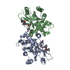
| ||||||||
| 2 | 
| ||||||||
| Unit cell |
| ||||||||
| Details | THERE ARE FOUR MOLECULES IN THE ASYMMETRIC UNIT, FORMING TWO NON-CRYSTALLOGRAPHIC DIMERS. |
-Components
| #1: Protein | Mass: 28178.676 Da / Num. of mol.: 4 / Mutation: A143Q, T181M, L229M Source method: isolated from a genetically manipulated source Source: (gene. exp.) Human herpesvirus 5 / Production host:  References: UniProt: P16753, Hydrolases; Acting on peptide bonds (peptidases); Serine endopeptidases #2: Chemical | ChemComp-01E / (   Type: peptide-like, Peptide-like / Class: Inhibitor / Mass: 709.615 Da / Num. of mol.: 4 / Source method: obtained synthetically / Formula: C31H44IN5O6 / References: BILC 821 Type: peptide-like, Peptide-like / Class: Inhibitor / Mass: 709.615 Da / Num. of mol.: 4 / Source method: obtained synthetically / Formula: C31H44IN5O6 / References: BILC 821Has protein modification | Y | |
|---|
-Experimental details
-Experiment
| Experiment | Method: X-RAY DIFFRACTION / Number of used crystals: 1 |
|---|
- Sample preparation
| Crystal | Density Matthews: 2.71 Å3/Da / Density % sol: 54.62 % | |||||||||||||||||||||||||||||||||||||||||||||||||
|---|---|---|---|---|---|---|---|---|---|---|---|---|---|---|---|---|---|---|---|---|---|---|---|---|---|---|---|---|---|---|---|---|---|---|---|---|---|---|---|---|---|---|---|---|---|---|---|---|---|---|
| Crystal grow | pH: 7.5 / Details: pH 7.5 | |||||||||||||||||||||||||||||||||||||||||||||||||
| Crystal grow | *PLUS Method: vapor diffusion, hanging drop | |||||||||||||||||||||||||||||||||||||||||||||||||
| Components of the solutions | *PLUS
|
-Data collection
| Diffraction | Mean temperature: 100 K |
|---|---|
| Diffraction source | Wavelength: 1.5418 |
| Radiation | Protocol: SINGLE WAVELENGTH / Monochromatic (M) / Laue (L): M / Scattering type: x-ray |
| Radiation wavelength | Wavelength: 1.5418 Å / Relative weight: 1 |
| Reflection | Resolution: 2.7→20 Å / Num. obs: 33502 / % possible obs: 93 % / Observed criterion σ(I): 1 / Redundancy: 4 % / Rmerge(I) obs: 0.078 |
| Reflection | *PLUS Num. measured all: 275527 |
- Processing
| Software |
| ||||||||||||||||||||||||||||||||||||||||||||||||||||||||||||
|---|---|---|---|---|---|---|---|---|---|---|---|---|---|---|---|---|---|---|---|---|---|---|---|---|---|---|---|---|---|---|---|---|---|---|---|---|---|---|---|---|---|---|---|---|---|---|---|---|---|---|---|---|---|---|---|---|---|---|---|---|---|
| Refinement | Method to determine structure: MOLECULAR REPLACEMENT Starting model: PDB ENTRY 1WPO Resolution: 2.7→6 Å / Cross valid method: THROUGHOUT / σ(F): 2
| ||||||||||||||||||||||||||||||||||||||||||||||||||||||||||||
| Refinement step | Cycle: LAST / Resolution: 2.7→6 Å
| ||||||||||||||||||||||||||||||||||||||||||||||||||||||||||||
| Refine LS restraints |
| ||||||||||||||||||||||||||||||||||||||||||||||||||||||||||||
| Refine LS restraints NCS | NCS model details: RESTRAINTS |
