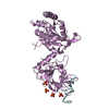
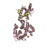
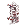
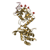
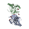
THERE ARE TWO MOLECULES IN THE ASYMMETRIC UNIT, FORMING A NON-CRYSTALLOGRAPHIC DIMER. THE RESIDUES IN THE FIRST MOLECULE ARE NUMBERED 1 A - 256 A, AND THOSE IN THE SECOND MOLECULE ARE NUMBERED 1 B - 256 B.
-
Components
#1: Protein
HUMANCYTOMEGALOVIRUSPROTEASE
Mass: 28262.309 Da / Num. of mol.: 2 Source method: isolated from a genetically manipulated source Source: (gene. exp.) Human herpesvirus 5 / Genus: Cytomegalovirus / Production host: Escherichia coli (E. coli) References: UniProt: P16753, Hydrolases; Acting on peptide bonds (peptidases); Serine endopeptidases
Mass: 18.015 Da / Num. of mol.: 249 / Source method: isolated from a natural source / Formula: H2O
Compound details
THE PROTEASE USED HERE CONTAIN THREE MUTATIONS: A143Q, T181M, AND L229M. IN ADDITION, THE PROTEASE ...THE PROTEASE USED HERE CONTAIN THREE MUTATIONS: A143Q, T181M, AND L229M. IN ADDITION, THE PROTEASE CONTAINS SELENO-METHIONYL RESIDUES (CALLED MSE) INSTEAD OF REGULAR METHIONYL RESIDUES.
Has protein modification
Y
-
Experimental details
-
Experiment
Experiment
Method: X-RAY DIFFRACTION
-
Sample preparation
Crystal
Density Matthews: 2.33 Å3/Da / Density % sol: 47.26 %
Crystal grow
*PLUS
pH: 5 / Method: unknown
Components of the solutions
*PLUS
ID
Conc.
Common name
Crystal-ID
Sol-ID
Chemical formula
1
25mg/ml
protein
1
1
2
20mM
sodiumacetate
1
1
3
80mM
1
1
NaCl
4
1mM
dithiothreitol
1
1
5
16 %
PEG4000
1
2
6
0.1M
MES
1
2
7
0.4M
1
2
LiCl
8
10 %
glycerol
1
2
9
5 %
t-butanol
1
2
-
Data collection
Radiation
Scattering type: x-ray
Radiation wavelength
Relative weight: 1
-
Processing
Software
Name
Classification
X-PLOR
modelbuilding
X-PLOR
refinement
X-PLOR
phasing
Refinement
Resolution: 2→6 Å / σ(F): 1
Rfactor
Num. reflection
% reflection
Rfree
0.288
-
7.5 %
Rwork
0.221
-
-
obs
0.221
30822
-
Refinement step
Cycle: LAST / Resolution: 2→6 Å
Protein
Nucleic acid
Ligand
Solvent
Total
Num. atoms
3285
0
5
249
3539
Refine LS restraints
Refine-ID
Type
Dev ideal
X-RAY DIFFRACTION
x_bond_d
0.01
X-RAY DIFFRACTION
x_bond_d_na
X-RAY DIFFRACTION
x_bond_d_prot
X-RAY DIFFRACTION
x_angle_d
X-RAY DIFFRACTION
x_angle_d_na
X-RAY DIFFRACTION
x_angle_d_prot
X-RAY DIFFRACTION
x_angle_deg
1.8
X-RAY DIFFRACTION
x_angle_deg_na
X-RAY DIFFRACTION
x_angle_deg_prot
X-RAY DIFFRACTION
x_dihedral_angle_d
24.2
X-RAY DIFFRACTION
x_dihedral_angle_d_na
X-RAY DIFFRACTION
x_dihedral_angle_d_prot
X-RAY DIFFRACTION
x_improper_angle_d
1.6
X-RAY DIFFRACTION
x_improper_angle_d_na
X-RAY DIFFRACTION
x_improper_angle_d_prot
X-RAY DIFFRACTION
x_mcbond_it
X-RAY DIFFRACTION
x_mcangle_it
X-RAY DIFFRACTION
x_scbond_it
X-RAY DIFFRACTION
x_scangle_it
Refine LS restraints
*PLUS
Refine-ID
Type
Dev ideal
X-RAY DIFFRACTION
x_dihedral_angle_d
X-RAY DIFFRACTION
x_dihedral_angle_deg
24.2
X-RAY DIFFRACTION
x_improper_angle_d
X-RAY DIFFRACTION
x_improper_angle_deg
1.6
+
About Yorodumi
-
News
-
Feb 9, 2022. New format data for meta-information of EMDB entries
New format data for meta-information of EMDB entries
Version 3 of the EMDB header file is now the official format.
The previous official version 1.9 will be removed from the archive.
In the structure databanks used in Yorodumi, some data are registered as the other names, "COVID-19 virus" and "2019-nCoV". Here are the details of the virus and the list of structure data.
Jan 31, 2019. EMDB accession codes are about to change! (news from PDBe EMDB page)
EMDB accession codes are about to change! (news from PDBe EMDB page)
The allocation of 4 digits for EMDB accession codes will soon come to an end. Whilst these codes will remain in use, new EMDB accession codes will include an additional digit and will expand incrementally as the available range of codes is exhausted. The current 4-digit format prefixed with “EMD-” (i.e. EMD-XXXX) will advance to a 5-digit format (i.e. EMD-XXXXX), and so on. It is currently estimated that the 4-digit codes will be depleted around Spring 2019, at which point the 5-digit format will come into force.
The EM Navigator/Yorodumi systems omit the EMD- prefix.
Related info.:Q: What is EMD? / ID/Accession-code notation in Yorodumi/EM Navigator
Yorodumi is a browser for structure data from EMDB, PDB, SASBDB, etc.
This page is also the successor to EM Navigator detail page, and also detail information page/front-end page for Omokage search.
The word "yorodu" (or yorozu) is an old Japanese word meaning "ten thousand". "mi" (miru) is to see.
Related info.:EMDB / PDB / SASBDB / Comparison of 3 databanks / Yorodumi Search / Aug 31, 2016. New EM Navigator & Yorodumi / Yorodumi Papers / Jmol/JSmol / Function and homology information / Changes in new EM Navigator and Yorodumi
 Movie
Movie Controller


 Open data
 Basic information
 Components
 Keywords
 Function and homology information
 Human herpesvirus 5
 X-RAY DIFFRACTION / Resolution: 2 Å
  Authors
 Citation
 Structure visualization
 Downloads & links
 Other downloads


 PDBj
PDBj Assembly


 Mass: 96.063 Da / Num. of mol.: 1 / Source method: obtained synthetically / Formula: SO4
 Mass: 18.015 Da / Num. of mol.: 249 / Source method: isolated from a natural source / Formula: H2O
 Sample preparation
 Processing