 Movie
Movie Controller

[English] 日本語
 Yorodumi
- PDB-2yep: STRUCTURE OF AN N-TERMINAL NUCLEOPHILE (NTN) HYDROLASE, OAT2, IN ... -
+ Open data

- Basic information
| Entry | Database: PDB / ID: 2yep | |||||||||||||||
|---|---|---|---|---|---|---|---|---|---|---|---|---|---|---|---|---|
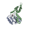
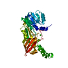
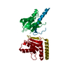
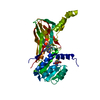
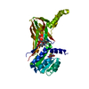
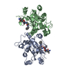
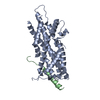
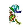
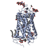
| Title | STRUCTURE OF AN N-TERMINAL NUCLEOPHILE (NTN) HYDROLASE, OAT2, IN COMPLEX WITH GLUTAMATE | |||||||||||||||
 Components Components |
| |||||||||||||||
 Keywords Keywords | TRANSFERASE / ACYL ENZYME / NTN HYDROLASE / ACYLTRANSFERASE / ORNITHINE ACETYL TRANSFERASE / HYDROLASE | |||||||||||||||
| Function / homology |  Function and homology information Function and homology informationL-methionine N-acyltransferase activity / glutamate N-acetyltransferase / L-glutamate N-acetyltransferase activity, acting on acetyl-L-ornithine as donor / L-ornithine biosynthetic process / amino-acid N-acetyltransferase / clavulanic acid biosynthetic process / L-glutamate N-acetyltransferase activity, acting on acetyl-CoA as donor / L-arginine biosynthetic process / cytoplasm Similarity search - Function | |||||||||||||||
| Biological species |  STREPTOMYCES CLAVULIGERUS (bacteria) STREPTOMYCES CLAVULIGERUS (bacteria) | |||||||||||||||
| Method |  X-RAY DIFFRACTION / MOLECULAR REPLACEMENT / Resolution: 2.7 Å X-RAY DIFFRACTION / MOLECULAR REPLACEMENT / Resolution: 2.7 Å | |||||||||||||||
 Authors Authors | Chowdhury, R. / Iqbal, A. / Clifton, I.J. / Schofield, C.J. | |||||||||||||||
 Citation Citation | Journal: Org.Biomol.Chem. / Year: 2011 Title: Structural and Biochemical Analyses Reveal How Ornithine Acetyl Transferase Binds Acidic and Basic Amino Acid Substrates. Authors: Iqbal, A. / Clifton, I.J. / Chowdhury, R. / Ivison, D. / Domene, C. / Schofield, C.J. | |||||||||||||||
| History |
|
- Structure visualization
| Structure viewer | Molecule: MolmilJmol/JSmol |
|---|
- Downloads & links
-Download
| PDBx/mmCIF format | 2yep.cif.gz | 290.6 KB | Display | PDBx/mmCIF format |
|---|---|---|---|---|
| PDB format | pdb2yep.ent.gz | 235.3 KB | Display | PDB format |
| PDBx/mmJSON format | 2yep.json.gz | Tree view | PDBx/mmJSON format | |
| Others |  Other downloads Other downloads |
-Validation report
| Arichive directory | https://data.pdbj.org/pub/pdb/validation_reports/ye/2yepftp://data.pdbj.org/pub/pdb/validation_reports/ye/2yep | HTTPS FTP |
|---|
-Related structure data
| Related structure data |  1vz6S S: Starting model for refinement |
|---|---|
| Similar structure data |


-Links
 PDBj
PDBj

- Assembly
| Deposited unit | 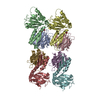
| ||||||||||||||||||||||||||||
|---|---|---|---|---|---|---|---|---|---|---|---|---|---|---|---|---|---|---|---|---|---|---|---|---|---|---|---|---|---|
| 1 | 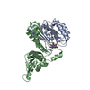
| ||||||||||||||||||||||||||||
| 2 | 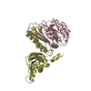
| ||||||||||||||||||||||||||||
| 3 | 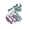
| ||||||||||||||||||||||||||||
| 4 | 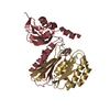
| ||||||||||||||||||||||||||||
| Unit cell |
| ||||||||||||||||||||||||||||
| Noncrystallographic symmetry (NCS) | NCS oper:
|
-Components
-Protein , 1 types, 4 molecules ACEG
| #1: Protein | Mass: 18837.371 Da / Num. of mol.: 4 Source method: isolated from a genetically manipulated source Source: (gene. exp.) STREPTOMYCES CLAVULIGERUS (bacteria) / Plasmid: PTYB12 / Production host: References: UniProt: Q53940, UniProt: P0DJQ5*PLUS, glutamate N-acetyltransferase |
|---|
-GLUTAMATE N-ACETYLTRANSFERASE 2 BETA ... , 2 types, 4 molecules BDHF
| #2: Protein | Mass: 22876.357 Da / Num. of mol.: 3 Source method: isolated from a genetically manipulated source Source: (gene. exp.) STREPTOMYCES CLAVULIGERUS (bacteria) / Plasmid: PTYB12 / Production host: References: UniProt: Q53940, UniProt: P0DJQ5*PLUS, glutamate N-acetyltransferase #3: Protein | | Mass: 22834.320 Da / Num. of mol.: 1 Source method: isolated from a genetically manipulated source Source: (gene. exp.) STREPTOMYCES CLAVULIGERUS (bacteria) / Plasmid: PTYB12 / Production host: References: UniProt: Q53940, UniProt: P0DJQ5*PLUS, glutamate N-acetyltransferase |
|---|
-Non-polymers , 3 types, 423 molecules 


| #4: Chemical |  Type: L-peptide linking / Mass: 147.129 Da / Num. of mol.: 2 / Source method: obtained synthetically / Formula: C5H9NO4 Type: L-peptide linking / Mass: 147.129 Da / Num. of mol.: 2 / Source method: obtained synthetically / Formula: C5H9NO4#5: Chemical |  Mass: 59.044 Da / Num. of mol.: 2 / Source method: obtained synthetically / Formula: C2H3O2 Mass: 59.044 Da / Num. of mol.: 2 / Source method: obtained synthetically / Formula: C2H3O2#6: Water | ChemComp-HOH / | Mass: 18.015 Da / Num. of mol.: 419 / Source method: isolated from a natural source / Formula: H2O |
|---|
-Details
| Has protein modification | Y |
|---|
-Experimental details
-Experiment
| Experiment | Method: X-RAY DIFFRACTION / Number of used crystals: 1 |
|---|
- Sample preparation
| Crystal | Density Matthews: 2.43 Å3/Da / Density % sol: 49.4 % Description: RMERGE AND REDUNDANCY VALUES NOT KNOWN, ESTIMATES GIVEN |
|---|---|
| Crystal grow | Temperature: 290 K / pH: 7.5 Details: 1.4M AMMONIUM SULPHATE, 100MM N-ACETYL -L-GLUTAMATE, 200MM NACL, 100MM TRIS HCL PH 7.5 . |
-Data collection
| Diffraction | Mean temperature: 100 K |
|---|---|
| Diffraction source | Source: SEALED TUBE / Type: OXFORD DIFFRACTION NOVA / Wavelength: 1.5418 / Wavelength: 1.5418 Å |
| Detector | Type: OXFORD DIFFRACTION / Detector: CCD / Date: Nov 10, 2006 / Details: MULTI-LAYER OPTIC |
| Radiation | Protocol: SINGLE WAVELENGTH / Monochromatic (M) / Laue (L): M / Scattering type: x-ray |
| Radiation wavelength | Wavelength: 1.5418 Å / Relative weight: 1 |
| Reflection | Resolution: 2.7→61.12 Å / Num. obs: 60743 / % possible obs: 96 % / Observed criterion σ(I): 0 / Redundancy: 1 % / Biso Wilson estimate: 14.5 Å2 / Rmerge(I) obs: 0.1 |
| Reflection shell | % possible all: 95.7 |
- Processing
| Software |
| ||||||||||||||||||||||||||||||||||||||||||||||||||||||||||||
|---|---|---|---|---|---|---|---|---|---|---|---|---|---|---|---|---|---|---|---|---|---|---|---|---|---|---|---|---|---|---|---|---|---|---|---|---|---|---|---|---|---|---|---|---|---|---|---|---|---|---|---|---|---|---|---|---|---|---|---|---|---|
| Refinement | Method to determine structure: MOLECULAR REPLACEMENT Starting model: PDB ENTRY 1VZ6, CHAIN A Resolution: 2.7→61.12 Å / Rfactor Rfree error: 0.005 / Data cutoff high absF: 3620114.47 / Data cutoff low absF: 0 / Isotropic thermal model: RESTRAINED / Cross valid method: THROUGHOUT / σ(F): 0 / Stereochemistry target values: MAXIMUM LIKELIHOOD / Details: HYDROGENS HAVE BEEN ADDED IN THE RIDING POSITIONS.
| ||||||||||||||||||||||||||||||||||||||||||||||||||||||||||||
| Solvent computation | Solvent model: FLAT MODEL / Bsol: 25.54 Å2 / ksol: 0.37 e/Å3 | ||||||||||||||||||||||||||||||||||||||||||||||||||||||||||||
| Displacement parameters | Biso mean: 12.8 Å2
| ||||||||||||||||||||||||||||||||||||||||||||||||||||||||||||
| Refine analyze |
| ||||||||||||||||||||||||||||||||||||||||||||||||||||||||||||
| Refinement step | Cycle: LAST / Resolution: 2.7→61.12 Å
| ||||||||||||||||||||||||||||||||||||||||||||||||||||||||||||
| Refine LS restraints |
| ||||||||||||||||||||||||||||||||||||||||||||||||||||||||||||
| Refine LS restraints NCS | NCS model details: NONE | ||||||||||||||||||||||||||||||||||||||||||||||||||||||||||||
| LS refinement shell | Resolution: 2.7→2.77 Å / Rfactor Rfree error: 0.019 / Total num. of bins used: 13
| ||||||||||||||||||||||||||||||||||||||||||||||||||||||||||||
| Xplor file |
|
