 Movie
Movie Controller

[English] 日本語
 Yorodumi
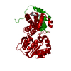
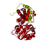
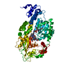
- PDB-1jdj: CRYSTAL STRUCTURE OF LEISHMANIA MEXICANA GLYCEROL-3-PHOSPHATE DEH... -
+ Open data

- Basic information
| Entry | Database: PDB / ID: 1jdj | ||||||
|---|---|---|---|---|---|---|---|
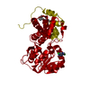
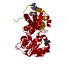
| Title | CRYSTAL STRUCTURE OF LEISHMANIA MEXICANA GLYCEROL-3-PHOSPHATE DEHYDROGENASE IN COMPLEX WITH 2-FLUORO-6-CHLOROPURINE | ||||||
 Components Components | GLYCEROL-3-PHOSPHATE DEHYDROGENASE | ||||||
 Keywords Keywords | OXIDOREDUCTASE / dehydrogenase | ||||||
| Function / homology |  Function and homology information Function and homology informationglycerol-3-phosphate dehydrogenase (NAD+) / glycerol-3-phosphate dehydrogenase (NAD+) activity / glycerol-3-phosphate catabolic process / glycosome / NAD binding / carbohydrate metabolic process / cytosol Similarity search - Function | ||||||
| Biological species |   Leishmania mexicana (eukaryote) Leishmania mexicana (eukaryote) | ||||||
| Method |  X-RAY DIFFRACTION / SYNCHROTRON / MOLECULAR REPLACEMENT / Resolution: 2.2 Å X-RAY DIFFRACTION / SYNCHROTRON / MOLECULAR REPLACEMENT / Resolution: 2.2 Å | ||||||
 Authors Authors | Suresh, S. / Wisedchaisri, G. / Kennedy, K.J. / Verlinde, C.L.M.J. / Gelb, M.H. / Hol, W.G.J. | ||||||
 Citation Citation | Journal: Chem.Biol. / Year: 2002 Title: Anomalous differences of light elements in determining precise binding modes of ligands to glycerol-3-phosphate dehydrogenase. Authors: Choe, J. / Suresh, S. / Wisedchaisri, G. / Kennedy, K.J. / Gelb, M.H. / Hol, W.G. | ||||||
| History |
|
- Structure visualization
| Structure viewer | Molecule: MolmilJmol/JSmol |
|---|
- Downloads & links
-Download
| PDBx/mmCIF format | 1jdj.cif.gz | 82 KB | Display | PDBx/mmCIF format |
|---|---|---|---|---|
| PDB format | pdb1jdj.ent.gz | 60.8 KB | Display | PDB format |
| PDBx/mmJSON format | 1jdj.json.gz | Tree view | PDBx/mmJSON format | |
| Others |  Other downloads Other downloads |
-Validation report
| Arichive directory | https://data.pdbj.org/pub/pdb/validation_reports/jd/1jdjftp://data.pdbj.org/pub/pdb/validation_reports/jd/1jdj | HTTPS FTP |
|---|
-Related structure data
| Related structure data |  1m66C 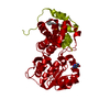 1m67C  1n1gC  1evyS S: Starting model for refinement C: citing same article ( |
|---|---|
| Similar structure data |


-Links
 PDBj
PDBj


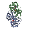
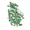
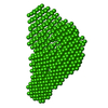
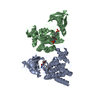
- Assembly
| Deposited unit | 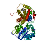
| ||||||||
|---|---|---|---|---|---|---|---|---|---|
| 1 | 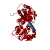
| ||||||||
| Unit cell |
| ||||||||
| Details | biological dimer generated by crystallographic two-fold |
-Components
| #1: Protein | Mass: 39317.828 Da / Num. of mol.: 1 Source method: isolated from a genetically manipulated source Source: (gene. exp.) Leishmania mexicana (eukaryote) / Plasmid: pET3a / Species (production host): Escherichia coli / Production host:  References: UniProt: P90551, glycerol-3-phosphate dehydrogenase (NAD+) |
|---|---|
| #2: Chemical | ChemComp-CFP / 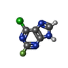  Mass: 172.548 Da / Num. of mol.: 1 / Source method: obtained synthetically / Formula: C5H2ClFN4 Mass: 172.548 Da / Num. of mol.: 1 / Source method: obtained synthetically / Formula: C5H2ClFN4 |
| #3: Chemical | ChemComp-MYS /   Mass: 212.415 Da / Num. of mol.: 1 / Source method: obtained synthetically / Formula: C15H32 Mass: 212.415 Da / Num. of mol.: 1 / Source method: obtained synthetically / Formula: C15H32 |
| #4: Water | ChemComp-HOH /  Mass: 18.015 Da / Num. of mol.: 160 / Source method: isolated from a natural source / Formula: H2O Mass: 18.015 Da / Num. of mol.: 160 / Source method: isolated from a natural source / Formula: H2O |
-Experimental details
-Experiment
| Experiment | Method: X-RAY DIFFRACTION / Number of used crystals: 1 |
|---|
- Sample preparation
| Crystal | Density Matthews: 3.31 Å3/Da / Density % sol: 62.87 % |
|---|---|
| Crystal grow | Temperature: 298 K / Method: vapor diffusion, sitting drop / pH: 7.2 Details: 0.9 M sodium citrate, 50 mM TEA pH 7.2, VAPOR DIFFUSION, SITTING DROP, temperature 298K |
| Crystal grow | *PLUS |
| Components of the solutions | *PLUS Conc.: 0.8-0.9 M / Common name: sodium citrate |
-Data collection
| Diffraction | Mean temperature: 125 K |
|---|---|
| Diffraction source | Source: SYNCHROTRON / Site: APS  / Beamline: 19-ID / Wavelength: 0.979 Å / Beamline: 19-ID / Wavelength: 0.979 Å |
| Detector | Type: CUSTOM-MADE / Detector: CCD / Date: Mar 1, 2001 |
| Radiation | Protocol: SINGLE WAVELENGTH / Monochromatic (M) / Laue (L): M / Scattering type: x-ray |
| Radiation wavelength | Wavelength: 0.979 Å / Relative weight: 1 |
| Reflection | Resolution: 2.2→50 Å / Num. all: 27686 / Num. obs: 27686 / % possible obs: 99.1 % / Observed criterion σ(F): 0 / Observed criterion σ(I): 0 / Redundancy: 7.08 % / Biso Wilson estimate: 28.44 Å2 / Rmerge(I) obs: 0.064 / Rsym value: 0.064 / Net I/σ(I): 27 |
| Reflection shell | Resolution: 2.2→2.28 Å / Redundancy: 7 % / Rmerge(I) obs: 0.184 / Mean I/σ(I) obs: 7.6 / Num. unique all: 2492 / Rsym value: 0.184 / % possible all: 91.6 |
| Reflection | *PLUS Redundancy: 7.1 % / Num. measured all: 196124 |
| Reflection shell | *PLUS % possible obs: 91.6 % / Rmerge(I) obs: 0.18 |
- Processing
| Software |
| |||||||||||||||||||||||||
|---|---|---|---|---|---|---|---|---|---|---|---|---|---|---|---|---|---|---|---|---|---|---|---|---|---|---|
| Refinement | Method to determine structure: MOLECULAR REPLACEMENT Starting model: PDB ENTRY 1EVY Resolution: 2.2→50 Å / Isotropic thermal model: isotropic / Cross valid method: THROUGHOUT / σ(F): 0 / σ(I): 0 / Stereochemistry target values: CNS / Details: CNS
| |||||||||||||||||||||||||
| Displacement parameters | Biso mean: 31.38 Å2
| |||||||||||||||||||||||||
| Refinement step | Cycle: LAST / Resolution: 2.2→50 Å
| |||||||||||||||||||||||||
| Refine LS restraints |
| |||||||||||||||||||||||||
| LS refinement shell | Resolution: 2.2→2.28 Å / Rfactor Rfree error: 0
| |||||||||||||||||||||||||
| Refinement | *PLUS | |||||||||||||||||||||||||
| Solvent computation | *PLUS | |||||||||||||||||||||||||
| Displacement parameters | *PLUS | |||||||||||||||||||||||||
| LS refinement shell | *PLUS Highest resolution: 2.2 Å / Rfactor obs: 0.215 |
