 Movie
Movie Controller


+ Open data

- Basic information
| Entry | Database: PDB / ID: 1k98 | ||||||
|---|---|---|---|---|---|---|---|
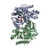
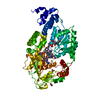
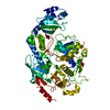
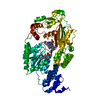
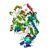
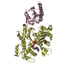
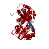
| Title | AdoMet complex of MetH C-terminal fragment | ||||||
 Components Components | Methionine synthase | ||||||
 Keywords Keywords | TRANSFERASE / AdoMet binding / MOTION OF 4-HELIX BUNDLE / DOMAIN INTERACTIONS | ||||||
| Function / homology |  Function and homology information Function and homology informationmethionine synthase / methionine synthase activity / homocysteine metabolic process / tetrahydrofolate metabolic process / cobalamin binding / : / tetrahydrofolate interconversion / methylation / zinc ion binding / cytosol / cytoplasm Similarity search - Function | ||||||
| Biological species |  | ||||||
| Method |  X-RAY DIFFRACTION / MOLECULAR REPLACEMENT / Resolution: 3.75 Å X-RAY DIFFRACTION / MOLECULAR REPLACEMENT / Resolution: 3.75 Å | ||||||
 Authors Authors | Bandarian, V. / Pattridge, K.A. / Lennon, B.W. / Huddler, D.P. / Matthews, R.G. / Ludwig, M.L. | ||||||
 Citation Citation | Journal: Nat.Struct.Biol. / Year: 2002 Title: Domain alternation switches B(12)-dependent methionine synthase to the activation conformation. Authors: Bandarian, V. / Pattridge, K.A. / Lennon, B.W. / Huddler, D.P. / Matthews, R.G. / Ludwig, M.L. | ||||||
| History |
|
- Structure visualization
| Structure viewer | Molecule: MolmilJmol/JSmol |
|---|
- Downloads & links
-Download
| PDBx/mmCIF format | 1k98.cif.gz | 131.2 KB | Display | PDBx/mmCIF format |
|---|---|---|---|---|
| PDB format | pdb1k98.ent.gz | 100.2 KB | Display | PDB format |
| PDBx/mmJSON format | 1k98.json.gz | Tree view | PDBx/mmJSON format | |
| Others |  Other downloads Other downloads |
-Validation report
| Arichive directory | https://data.pdbj.org/pub/pdb/validation_reports/k9/1k98ftp://data.pdbj.org/pub/pdb/validation_reports/k9/1k98 | HTTPS FTP |
|---|
-Related structure data
| Related structure data | 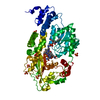 1k7ySC S: Starting model for refinement C: citing same article ( |
|---|---|
| Similar structure data |


-Links
 PDBj
PDBj- Assembly
| Deposited unit | 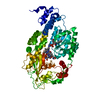
| ||||||||
|---|---|---|---|---|---|---|---|---|---|
| 1 |
| ||||||||
| Unit cell |
|
-Components
| #1: Protein | Mass: 64859.137 Da / Num. of mol.: 1 / Fragment: c-terminal activation complex, residues 651-1227 / Mutation: H759G Source method: isolated from a genetically manipulated source Source: (gene. exp.) |
|---|---|
| #2: Chemical | ChemComp-SO4 /   Mass: 96.063 Da / Num. of mol.: 1 / Source method: obtained synthetically / Formula: SO4 Mass: 96.063 Da / Num. of mol.: 1 / Source method: obtained synthetically / Formula: SO4 |
| #3: Chemical | ChemComp-B12 /   Mass: 1330.356 Da / Num. of mol.: 1 / Source method: obtained synthetically / Formula: C62H89CoN13O14P Mass: 1330.356 Da / Num. of mol.: 1 / Source method: obtained synthetically / Formula: C62H89CoN13O14P |
-Experimental details
-Experiment
| Experiment | Method: X-RAY DIFFRACTION / Number of used crystals: 1 |
|---|
- Sample preparation
| Crystal | Density Matthews: 3.32 Å3/Da / Density % sol: 62.97 % | ||||||||||||||||||||||||||||||
|---|---|---|---|---|---|---|---|---|---|---|---|---|---|---|---|---|---|---|---|---|---|---|---|---|---|---|---|---|---|---|---|
| Crystal grow | Temperature: 295 K / Method: vapor diffusion, hanging drop / pH: 6.5 Details: cacodylate, ammonium sulfate, PEG 8000, pH 6.5, VAPOR DIFFUSION, HANGING DROP, temperature 295K | ||||||||||||||||||||||||||||||
| Crystal grow | *PLUS Temperature: 22 ℃ | ||||||||||||||||||||||||||||||
| Components of the solutions | *PLUS
|
-Data collection
| Diffraction | Mean temperature: 140 K |
|---|---|
| Diffraction source | Source: ROTATING ANODE / Type: RIGAKU / Wavelength: 1.54 Å |
| Detector | Type: RIGAKU RAXIS IV / Detector: IMAGE PLATE / Date: May 9, 2000 / Details: Yale mirrors |
| Radiation | Monochromator: Yale Mirrors / Protocol: SINGLE WAVELENGTH / Monochromatic (M) / Laue (L): M / Scattering type: x-ray |
| Radiation wavelength | Wavelength: 1.54 Å / Relative weight: 1 |
| Reflection | Resolution: 3.75→50 Å / Num. all: 8076 / Num. obs: 8076 / % possible obs: 86.8 % / Observed criterion σ(I): 0 / Rsym value: 0.063 |
| Reflection shell | Resolution: 3.75→3.88 Å / Rsym value: 0.451 / % possible all: 66.9 |
| Reflection | *PLUS Lowest resolution: 35 Å / Num. obs: 8057 / % possible obs: 85.1 % / Num. measured all: 34361 / Rmerge(I) obs: 0.062 |
| Reflection shell | *PLUS % possible obs: 62.8 % / Rmerge(I) obs: 0.444 |
- Processing
| Software |
| |||||||||||||||||||||||||
|---|---|---|---|---|---|---|---|---|---|---|---|---|---|---|---|---|---|---|---|---|---|---|---|---|---|---|
| Refinement | Method to determine structure: MOLECULAR REPLACEMENT Starting model: PDB entry 1k7y Resolution: 3.75→15 Å / Cross valid method: THROUGHOUT / σ(F): 0 / Stereochemistry target values: Engh & Huber
| |||||||||||||||||||||||||
| Refinement step | Cycle: LAST / Resolution: 3.75→15 Å
| |||||||||||||||||||||||||
| LS refinement shell | Resolution: 3.75→3.88 Å /
| |||||||||||||||||||||||||
| Software | *PLUS Name: CNS / Classification: refinement | |||||||||||||||||||||||||
| Refinement | *PLUS Lowest resolution: 15 Å / σ(F): 0 / Rfactor obs: 0.308 | |||||||||||||||||||||||||
| Solvent computation | *PLUS | |||||||||||||||||||||||||
| Displacement parameters | *PLUS | |||||||||||||||||||||||||
| Refine LS restraints | *PLUS
| |||||||||||||||||||||||||
| LS refinement shell | *PLUS Rfactor Rfree: 0.48 / Rfactor Rwork: 0.478 |
