 Movie
Movie Controller

[English] 日本語
 Yorodumi
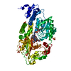
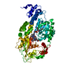
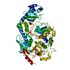
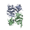
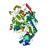
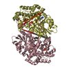
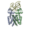
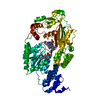
- PDB-3iva: Structure of the B12-dependent Methionine Synthase (MetH) C-temin... -
+ Open data

- Basic information
| Entry | Database: PDB / ID: 3iva | ||||||
|---|---|---|---|---|---|---|---|
| Title | Structure of the B12-dependent Methionine Synthase (MetH) C-teminal half with AdoHcy bound | ||||||
 Components Components | Methionine synthase | ||||||
 Keywords Keywords | TRANSFERASE / METH / reactivation conformation / H759 / cobalamin / intermodular interactions / amino-acid biosynthesis / cobalt / metal-binding / methionine biosynthesis / methyltransferase / S-adenosyl-L-methionine / S-adenosyl-homocysteine | ||||||
| Function / homology |  Function and homology information Function and homology informationmethionine synthase / methionine synthase activity / homocysteine metabolic process / cobalamin binding / tetrahydrofolate metabolic process / : / tetrahydrofolate interconversion / methylation / zinc ion binding / cytoplasm / cytosol Similarity search - Function | ||||||
| Biological species |  | ||||||
| Method |  X-RAY DIFFRACTION / SYNCHROTRON / MOLECULAR REPLACEMENT / Resolution: 2.7 Å X-RAY DIFFRACTION / SYNCHROTRON / MOLECULAR REPLACEMENT / Resolution: 2.7 Å | ||||||
 Authors Authors | Pattridge, K.A. / Koutmos, M. / Smith, J.L. | ||||||
 Citation Citation | Journal: Proc.Natl.Acad.Sci.USA / Year: 2009 Title: Insights into the reactivation of cobalamin-dependent methionine synthase. Authors: Koutmos, M. / Datta, S. / Pattridge, K.A. / Smith, J.L. / Matthews, R.G. | ||||||
| History |
|
- Structure visualization
| Structure viewer | Molecule: MolmilJmol/JSmol |
|---|
- Downloads & links
-Download
| PDBx/mmCIF format | 3iva.cif.gz | 134.6 KB | Display | PDBx/mmCIF format |
|---|---|---|---|---|
| PDB format | pdb3iva.ent.gz | 101.8 KB | Display | PDB format |
| PDBx/mmJSON format | 3iva.json.gz | Tree view | PDBx/mmJSON format | |
| Others |  Other downloads Other downloads |
-Validation report
| Arichive directory | https://data.pdbj.org/pub/pdb/validation_reports/iv/3ivaftp://data.pdbj.org/pub/pdb/validation_reports/iv/3iva | HTTPS FTP |
|---|
-Related structure data
| Related structure data |  3iv9C  3bulS S: Starting model for refinement C: citing same article ( |
|---|---|
| Similar structure data |


-Links
 PDBj
PDBj- Assembly
| Deposited unit | 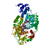
| ||||||||
|---|---|---|---|---|---|---|---|---|---|
| 1 |
| ||||||||
| Unit cell |
|
-Components
| #1: Protein | Mass: 65175.504 Da / Num. of mol.: 1 Fragment: C-terminal activation complex (UNP residues 649-1227) Mutation: I690C, G743C Source method: isolated from a genetically manipulated source Source: (gene. exp.) | ||||
|---|---|---|---|---|---|
| #2: Chemical | ChemComp-B12 /   Mass: 1330.356 Da / Num. of mol.: 1 / Source method: obtained synthetically / Formula: C62H89CoN13O14P Mass: 1330.356 Da / Num. of mol.: 1 / Source method: obtained synthetically / Formula: C62H89CoN13O14P | ||||
| #3: Chemical | ChemComp-SAH /   Mass: 384.411 Da / Num. of mol.: 1 / Source method: obtained synthetically / Formula: C14H20N6O5S Mass: 384.411 Da / Num. of mol.: 1 / Source method: obtained synthetically / Formula: C14H20N6O5S | ||||
| #4: Chemical | ChemComp-NO3 /   Mass: 62.005 Da / Num. of mol.: 4 / Source method: obtained synthetically / Formula: NO3 Mass: 62.005 Da / Num. of mol.: 4 / Source method: obtained synthetically / Formula: NO3#5: Water | ChemComp-HOH / |  Mass: 18.015 Da / Num. of mol.: 26 / Source method: isolated from a natural source / Formula: H2O Mass: 18.015 Da / Num. of mol.: 26 / Source method: isolated from a natural source / Formula: H2OHas protein modification | Y | |
-Experimental details
-Experiment
| Experiment | Method: X-RAY DIFFRACTION / Number of used crystals: 1 |
|---|
- Sample preparation
| Crystal | Density Matthews: 3.11 Å3/Da / Density % sol: 60.4 % |
|---|---|
| Crystal grow | Temperature: 302 K / Method: vapor diffusion / pH: 7 Details: 0.2 M potassium nitrate, 18 % (w/v) PEG3350, pH 7.0, VAPOR DIFFUSION, temperature 302K |
-Data collection
| Diffraction | Mean temperature: 100 K |
|---|---|
| Diffraction source | Source: SYNCHROTRON / Site: APS  / Beamline: 23-ID-B / Wavelength: 0.9793 Å / Beamline: 23-ID-B / Wavelength: 0.9793 Å |
| Detector | Type: MARMOSAIC 300 mm CCD / Detector: CCD / Date: Apr 2, 2007 Details: K-B pair of biomorph mirrors for vertical and horizontal focusing |
| Radiation | Monochromator: Double Crystal Monochromator / Protocol: SINGLE WAVELENGTH / Monochromatic (M) / Laue (L): M / Scattering type: x-ray |
| Radiation wavelength | Wavelength: 0.9793 Å / Relative weight: 1 |
| Reflection | Resolution: 2.7→50 Å / Num. all: 23160 / Num. obs: 23160 / % possible obs: 100 % / Observed criterion σ(F): 0 / Observed criterion σ(I): 0 / Redundancy: 22.4 % / Rmerge(I) obs: 0.043 / Rsym value: 0.085 / Net I/σ(I): 27.3 |
| Reflection shell | Resolution: 2.7→2.86 Å / Redundancy: 22.4 % / Rmerge(I) obs: 0.204 / Mean I/σ(I) obs: 5.82 / Rsym value: 0.622 / % possible all: 99.9 |
- Processing
| Software |
| ||||||||||||||||||||||||||||||||||||||||||||||||||||||||||||||||||||||||||||||||
|---|---|---|---|---|---|---|---|---|---|---|---|---|---|---|---|---|---|---|---|---|---|---|---|---|---|---|---|---|---|---|---|---|---|---|---|---|---|---|---|---|---|---|---|---|---|---|---|---|---|---|---|---|---|---|---|---|---|---|---|---|---|---|---|---|---|---|---|---|---|---|---|---|---|---|---|---|---|---|---|---|---|
| Refinement | Method to determine structure: MOLECULAR REPLACEMENT Starting model: Individual domains of PDB entry 3BUL Resolution: 2.7→45.32 Å / Rfactor Rfree error: 0.009 / Data cutoff high absF: 3333675.2 / Data cutoff low absF: 0 / Isotropic thermal model: RESTRAINED / Cross valid method: THROUGHOUT / σ(F): 0 / Stereochemistry target values: Engh & Huber / Details: BULK SOLVENT MODEL USED
| ||||||||||||||||||||||||||||||||||||||||||||||||||||||||||||||||||||||||||||||||
| Solvent computation | Solvent model: FLAT MODEL / Bsol: 60.9901 Å2 / ksol: 0.35 e/Å3 | ||||||||||||||||||||||||||||||||||||||||||||||||||||||||||||||||||||||||||||||||
| Displacement parameters | Biso mean: 76.7 Å2
| ||||||||||||||||||||||||||||||||||||||||||||||||||||||||||||||||||||||||||||||||
| Refine analyze |
| ||||||||||||||||||||||||||||||||||||||||||||||||||||||||||||||||||||||||||||||||
| Refinement step | Cycle: LAST / Resolution: 2.7→45.32 Å
| ||||||||||||||||||||||||||||||||||||||||||||||||||||||||||||||||||||||||||||||||
| Refine LS restraints |
| ||||||||||||||||||||||||||||||||||||||||||||||||||||||||||||||||||||||||||||||||
| Refine LS restraints NCS | NCS model details: NONE | ||||||||||||||||||||||||||||||||||||||||||||||||||||||||||||||||||||||||||||||||
| LS refinement shell | Resolution: 2.7→2.87 Å / Rfactor Rfree error: 0.029 / Total num. of bins used: 6
| ||||||||||||||||||||||||||||||||||||||||||||||||||||||||||||||||||||||||||||||||
| Xplor file |
|
