 Movie
Movie Controller
[English] 日本語
 Yorodumi
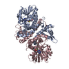
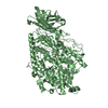
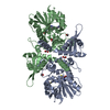
- SASDFC2: Urokinase plasminogen activator surface receptor, uPAR K50C-V70C,... -
+ Open data

- Basic information
| Entry | Database: SASBDB / ID: SASDFC2 |
|---|---|
 Sample Sample | Urokinase plasminogen activator surface receptor, uPAR K50C-V70C, complex with urokinase-type plasminogen activator (Amino Terminal Fragment, ATF).
|
| Function / homology |  Function and homology information Function and homology informationurokinase plasminogen activator receptor activity / Attachment of GPI anchor to uPAR / u-plasminogen activator / regulation of smooth muscle cell-matrix adhesion / urokinase plasminogen activator signaling pathway / regulation of plasminogen activation / positive regulation of homotypic cell-cell adhesion / regulation of integrin-mediated signaling pathway / regulation of fibrinolysis / protein complex involved in cell-matrix adhesion ...urokinase plasminogen activator receptor activity / Attachment of GPI anchor to uPAR / u-plasminogen activator / regulation of smooth muscle cell-matrix adhesion / urokinase plasminogen activator signaling pathway / regulation of plasminogen activation / positive regulation of homotypic cell-cell adhesion / regulation of integrin-mediated signaling pathway / regulation of fibrinolysis / protein complex involved in cell-matrix adhesion / regulation of wound healing / negative regulation of plasminogen activation / serine-type endopeptidase complex / regulation of smooth muscle cell migration / Dissolution of Fibrin Clot / regulation of cell adhesion mediated by integrin / extrinsic component of membrane / smooth muscle cell migration / positive regulation of DNA binding / plasminogen activation / negative regulation of intrinsic apoptotic signaling pathway / positive regulation of release of cytochrome c from mitochondria / tertiary granule membrane / regulation of proteolysis / negative regulation of fibrinolysis / regulation of cell adhesion / serine protease inhibitor complex / specific granule membrane / fibrinolysis / positive regulation of epidermal growth factor receptor signaling pathway / cell projection / positive regulation of protein phosphorylation / chemotaxis / blood coagulation / regulation of cell population proliferation / signaling receptor activity / response to hypoxia / positive regulation of cell migration / endoplasmic reticulum lumen / receptor ligand activity / serine-type endopeptidase activity / signaling receptor binding / external side of plasma membrane / protein domain specific binding / focal adhesion / Neutrophil degranulation / endoplasmic reticulum membrane / negative regulation of apoptotic process / enzyme binding / cell surface / signal transduction / proteolysis / : / extracellular exosome / extracellular region / membrane / plasma membrane Similarity search - Function |
| Biological species |  Homo sapiens (human) Homo sapiens (human) |
 Citation Citation | Journal: J Biol Chem / Year: 2019 Title: Did evolution create a flexible ligand-binding cavity in the urokinase receptor through deletion of a plesiotypic disulfide bond? Authors: Julie M Leth / Haydyn D T Mertens / Katrine Zinck Leth-Espensen / Thomas J D Jørgensen / Michael Ploug /   Abstract: The urokinase receptor (uPAR) is a founding member of a small protein family with multiple Ly6/uPAR (LU) domains. The motif defining these LU domains contains five plesiotypic disulfide bonds ...The urokinase receptor (uPAR) is a founding member of a small protein family with multiple Ly6/uPAR (LU) domains. The motif defining these LU domains contains five plesiotypic disulfide bonds stabilizing its prototypical three-fingered fold having three protruding loops. Notwithstanding the detailed knowledge on structure-function relationships in uPAR, one puzzling enigma remains unexplored. Why does the first LU domain in uPAR (DI) lack one of its consensus disulfide bonds, when the absence of this particular disulfide bond impairs the correct folding of other single LU domain-containing proteins? Here, using a variety of contemporary biophysical methods, we found that reintroducing the two missing half-cystines in uPAR DI caused the spontaneous formation of the corresponding consensus 7-8 LU domain disulfide bond. Importantly, constraints due to this cross-link impaired (i) the binding of uPAR to its primary ligand urokinase and (ii) the flexible interdomain assembly of the three LU domains in uPAR. We conclude that the evolutionary deletion of this particular disulfide bond in uPAR DI may have enabled the assembly of a high-affinity urokinase-binding cavity involving all three LU domains in uPAR. Of note, an analogous neofunctionalization occurred in snake venom α-neurotoxins upon loss of another pair of the plesiotypic LU domain half-cystines. In summary, elimination of the 7-8 consensus disulfide bond in the first LU domain of uPAR have significant functional and structural consequences. |
 Contact author Contact author |
|
- Structure visualization
| Structure viewer | Molecule: MolmilJmol/JSmol |
|---|
- Downloads & links
-Data source
| SASBDB page |  SASDFC2 SASDFC2 |
|---|
-Related structure data
| Related structure data | C: citing same article ( |
|---|---|
| Similar structure data |


-External links
| Related items in Molecule of the Month |
|---|


-Models
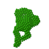
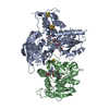
| Model #2811 |  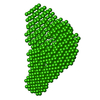 Type: dummy / Radius of dummy atoms: 3.00 A Comment: Refined DAMMIN model from average volume (10 DAMMIF iterations) Chi-square value: 1.433  Search similar-shape structures of this assembly by Omokage search (details) Search similar-shape structures of this assembly by Omokage search (details) |
|---|
-Sample
| Sample | Name: Urokinase plasminogen activator surface receptor, uPAR K50C-V70C, complex with urokinase-type plasminogen activator (Amino Terminal Fragment, ATF). Specimen concentration: 0.30-2.50 / Entity id: 66 / 1481 |
|---|---|
| Buffer | Name: 20 mM PBS, 5 %(v/v) glycerol / pH: 7.4 |
| Entity #66 | Name: uPAR / Type: protein Description: Urokinase plasminogen activator surface receptor Formula weight: 36.98 / Num. of mol.: 1 / Source: Homo sapiens / References: UniProt: Q03405 Sequence: MGHPPLLPLL LLLHTCVPAS WGLRCMQCKT NGDCRVEECA LGQDLCRTTI VRLWEEGEEL ELVEKSCTHS EKTNRTLSYR TGLKITSLTE VVCGLDLCNQ GNSGRAVTYS RSRYLECISC GSSDMSCERG RHQSLQCRSP EEQCLDVVTH WIQEGEEGRP KDDRHLRGCG ...Sequence: MGHPPLLPLL LLLHTCVPAS WGLRCMQCKT NGDCRVEECA LGQDLCRTTI VRLWEEGEEL ELVEKSCTHS EKTNRTLSYR TGLKITSLTE VVCGLDLCNQ GNSGRAVTYS RSRYLECISC GSSDMSCERG RHQSLQCRSP EEQCLDVVTH WIQEGEEGRP KDDRHLRGCG YLPGCPGSNG FHNNDTFHFL KCCNTTKCNE GPILELENLP QNGRQCYSCK GNSTHGCSSE ETFLIDCRGP MNQCLVATGT HEPKNQSYMV RGCATASMCQ HAHLGDAFSM NHIDVSCCTK SGCNHPDLDV QYRSGAAPQP GPAHLSLTIT LLMTARLWGG TLLWT |
| Entity #1481 | Name: ATF / Type: protein Description: Urokinase-type plasminogen activator (Amino Terminal Fragment) Formula weight: 16.099 / Num. of mol.: 1 / Source: Homo sapiens / References: UniProt: P00749 Sequence: MRALLARLLL CVLVVSDSKG SNELHQVPSN CDCLNGGTCV SNKYFSNIHW CNCPKKFGGQ HCEIDKSKTC YEGNGHFYRG KASTDTMGRP CLPWNSATVL QQTYHAHRSD ALQLGLGKHN YCRNPDNRRR PWCYVQVGLK PLV |
-Experimental information
| Beam | Instrument name: PETRA III EMBL P12 / City: Hamburg / 国: Germany / Type of source: X-ray synchrotron / Wavelength: 0.124 Å / Dist. spec. to detc.: 3.1 mm | |||||||||||||||||||||||||||||||||||||||
|---|---|---|---|---|---|---|---|---|---|---|---|---|---|---|---|---|---|---|---|---|---|---|---|---|---|---|---|---|---|---|---|---|---|---|---|---|---|---|---|---|
| Detector | Name: Pilatus 2M | |||||||||||||||||||||||||||||||||||||||
| Scan |  Measurement date: May 5, 2017 / Storage temperature: 10 °C / Cell temperature: 10 °C / Exposure time: 0.05 sec. / Number of frames: 20 / Unit: 1/nm /
| |||||||||||||||||||||||||||||||||||||||
| Distance distribution function P(R) | 
| |||||||||||||||||||||||||||||||||||||||
| Result |  Comments: Introduced disulfide in the first LU domain, DI, of uPAR (K50C-V70C).
|
