 Movie
Movie Controller

[English] 日本語
 Yorodumi
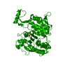
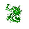
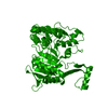
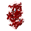
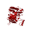
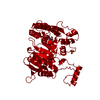
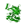
- PDB-1ide: ISOCITRATE DEHYDROGENASE Y160F MUTANT STEADY-STATE INTERMEDIATE C... -
+ Open data

- Basic information
| Entry | Database: PDB / ID: 1ide | ||||||
|---|---|---|---|---|---|---|---|
| Title | ISOCITRATE DEHYDROGENASE Y160F MUTANT STEADY-STATE INTERMEDIATE COMPLEX (LAUE DETERMINATION) | ||||||
 Components Components | ISOCITRATE DEHYDROGENASE | ||||||
 Keywords Keywords | OXIDOREDUCTASE (NAD(A)-CHOH(D)) | ||||||
| Function / homology |  Function and homology informationisocitrate dehydrogenase (NADP+) / isocitrate dehydrogenase (NADP+) activity / glyoxylate cycle / guanosine tetraphosphate binding / electron transport chain / tricarboxylic acid cycle / NAD binding / response to oxidative stress / magnesium ion binding / cytosol / cytoplasm Function and homology informationisocitrate dehydrogenase (NADP+) / isocitrate dehydrogenase (NADP+) activity / glyoxylate cycle / guanosine tetraphosphate binding / electron transport chain / tricarboxylic acid cycle / NAD binding / response to oxidative stress / magnesium ion binding / cytosol / cytoplasmSimilarity search - Function | ||||||
| Biological species |  Escherichia coli (E. coli) Escherichia coli (E. coli) | ||||||
| Method | X-RAY DIFFRACTION / SYNCHROTRON / Resolution: 2.5 Å | ||||||
 Authors Authors | Bolduc, J.M. / Dyer, D.H. / Scott, W.G. / Singer, P. / Sweet, R.M. / Koshland Junior, D.E. / Stoddard, B.L. | ||||||
 Citation Citation | Journal: Science / Year: 1995 Title: Mutagenesis and Laue structures of enzyme intermediates: isocitrate dehydrogenase. Authors: Bolduc, J.M. / Dyer, D.H. / Scott, W.G. / Singer, P. / Sweet, R.M. / Koshland Jr., D.E. / Stoddard, B.L. #1: Journal: Biochemistry / Year: 1991Title: Catalytic Mechanism of Nadp+-Dependent Isocitrate Dehydrogenase: Implications from the Structures of Magnesium-Isocitrate and Nadp+ Complexes Authors: Hurley, J.H. / Dean, A.M. / Koshland Junior, D.E. / Stroud, R.M. #2: Journal: J.Biol.Chem. / Year: 1990Title: Regulation of Isocitrate Dehydrogenase by Phosphorylation Involves No Long-Range Conformational Change in the Free Enzyme Authors: Hurley, J.H. / Dean, A.M. / Thorsness, P.E. / Koshland Junior, D.E. / Stroud, R.M. #3: Journal: Science / Year: 1990Title: Regulation of an Enzyme by Phosphorylation at the Active Site Authors: Hurley, J.H. / Dean, A.M. / Sohl, J.L. / Koshland Junior, D.E. / Stroud, R.M. #4: Journal: Proc.Natl.Acad.Sci.USA / Year: 1989Title: Structure of a Bacterial Enzyme Regulated by Phosphorylation, Isocitrate Dehydrogenase Authors: Hurley, J.H. / Thorsness, P.E. / Ramalingam, V. / Helmers, N.H. / Koshland Junior, D.E. / Stroud, R.M. | ||||||
| History |
|
- Structure visualization
| Structure viewer | Molecule: MolmilJmol/JSmol |
|---|
- Downloads & links
-Download
| PDBx/mmCIF format | 1ide.cif.gz | 109.8 KB | Display | PDBx/mmCIF format |
|---|---|---|---|---|
| PDB format | pdb1ide.ent.gz | 84.8 KB | Display | PDB format |
| PDBx/mmJSON format | 1ide.json.gz | Tree view | PDBx/mmJSON format | |
| Others |  Other downloads Other downloads |
-Validation report
| Arichive directory | https://data.pdbj.org/pub/pdb/validation_reports/id/1ideftp://data.pdbj.org/pub/pdb/validation_reports/id/1ide | HTTPS FTP |
|---|
-Related structure data


-Links
 PDBj
PDBj


- Assembly
| Deposited unit | 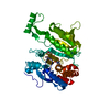
| ||||||||
|---|---|---|---|---|---|---|---|---|---|
| 1 | 
| ||||||||
| Unit cell |
| ||||||||
| Atom site foot note | 1: CIS PROLINE - PRO 262 |
-Components
| #1: Protein | / IDH Mass: 45793.562 Da / Num. of mol.: 1 / Mutation: Y160F Source method: isolated from a genetically manipulated source Details: TERNARY RATE-LIMITED MICHAELIS COMPLEX / Source: (gene. exp.) Escherichia coli (E. coli) / Strain: JLK1 / Gene: ICD / Variant: ICD(-) (DEFICIENT IN WT IDH GENE) / Plasmid: PTK513 / Gene (production host): ICDProduction host: PEMBL (DENTE ET AL 1983 NUC ACIDS RES 11,1645) References: UniProt: P08200, isocitrate dehydrogenase (NADP+) |
|---|---|
| #2: Chemical | ChemComp-MG /   Mass: 24.305 Da / Num. of mol.: 1 / Source method: obtained synthetically / Formula: Mg Mass: 24.305 Da / Num. of mol.: 1 / Source method: obtained synthetically / Formula: Mg |
| #3: Chemical | ChemComp-ICT / Isocitric acid  Mass: 192.124 Da / Num. of mol.: 1 / Source method: obtained synthetically / Formula: C6H8O7 Mass: 192.124 Da / Num. of mol.: 1 / Source method: obtained synthetically / Formula: C6H8O7 |
| #4: Chemical | ChemComp-NAP / Nicotinamide adenine dinucleotide phosphate  Mass: 743.405 Da / Num. of mol.: 1 / Source method: obtained synthetically / Formula: C21H28N7O17P3 Mass: 743.405 Da / Num. of mol.: 1 / Source method: obtained synthetically / Formula: C21H28N7O17P3 |
| #5: Water | ChemComp-HOH / Water Mass: 18.015 Da / Num. of mol.: 2 / Source method: isolated from a natural source / Formula: H2O Mass: 18.015 Da / Num. of mol.: 2 / Source method: isolated from a natural source / Formula: H2O |
| Compound details | THE NICOTINAMIDE RING IN THIS Y160F DYNAMIC LAUE STRUCTURE SHOWS VERY CLEAR SIGNS OF OCCUPYING AT ...THE NICOTINAMI |
-Experimental details
-Experiment
| Experiment | Method: X-RAY DIFFRACTION |
|---|
- Sample preparation
| Crystal | Density Matthews: 4.53 Å3/Da / Density % sol: 72.85 % | ||||||||||||||||||||||||||||||||||||||||||
|---|---|---|---|---|---|---|---|---|---|---|---|---|---|---|---|---|---|---|---|---|---|---|---|---|---|---|---|---|---|---|---|---|---|---|---|---|---|---|---|---|---|---|---|
| Crystal | *PLUS Density % sol: 73 % | ||||||||||||||||||||||||||||||||||||||||||
| Crystal grow | *PLUS pH: 5.4 / Method: unknown | ||||||||||||||||||||||||||||||||||||||||||
| Components of the solutions | *PLUS
|
-Data collection
| Diffraction source | Source: SYNCHROTRON / Site: NSLS  / Beamline: X26C / Wavelength: 0.7 - 2.1 / Beamline: X26C / Wavelength: 0.7 - 2.1 | |||||||||
|---|---|---|---|---|---|---|---|---|---|---|
| Detector | Type: FUJI / Detector: IMAGE PLATE / Date: Oct 15, 1993 | |||||||||
| Radiation | Protocol: LAUE / Monochromatic (M) / Laue (L): L / Scattering type: neutron | |||||||||
| Radiation wavelength |
| |||||||||
| Reflection | Num. obs: 17043 / % possible obs: 61 % / Observed criterion σ(I): 1 / Redundancy: 17.3 % / Rmerge(I) obs: 0.084 | |||||||||
| Reflection | *PLUS Highest resolution: 2.5 Å / Lowest resolution: 100 Å / Rmerge(I) obs: 0.084 |
- Processing
| Software |
| ||||||||||||||||||||||||||||||||||||||||||||||||||||||||||||
|---|---|---|---|---|---|---|---|---|---|---|---|---|---|---|---|---|---|---|---|---|---|---|---|---|---|---|---|---|---|---|---|---|---|---|---|---|---|---|---|---|---|---|---|---|---|---|---|---|---|---|---|---|---|---|---|---|---|---|---|---|---|
| Refinement | Highest resolution: 2.5 Å / σ(F): 2 Details: DATA COLLECTED FROM FOUR SEPARATE CRYSTALS AT X26-C (POLYCHROMATIC LAUE BEAM LINE AT BROOKHAVEN NATIONAL LABORATORY) AND MERGED TOGETHER WITH LAUENORM IN OXFORD LAUE DATA REDUCTION PACKAGE
| ||||||||||||||||||||||||||||||||||||||||||||||||||||||||||||
| Refinement step | Cycle: LAST / Highest resolution: 2.5 Å
| ||||||||||||||||||||||||||||||||||||||||||||||||||||||||||||
| Refine LS restraints |
| ||||||||||||||||||||||||||||||||||||||||||||||||||||||||||||
| Software | *PLUS Name: X-PLOR / Classification: refinement | ||||||||||||||||||||||||||||||||||||||||||||||||||||||||||||
| Refinement | *PLUS Lowest resolution: 10 Å | ||||||||||||||||||||||||||||||||||||||||||||||||||||||||||||
| Solvent computation | *PLUS | ||||||||||||||||||||||||||||||||||||||||||||||||||||||||||||
| Displacement parameters | *PLUS | ||||||||||||||||||||||||||||||||||||||||||||||||||||||||||||
| Refine LS restraints | *PLUS
|
