 Movie
Movie Controller

[English] 日本語
 Yorodumi
- PDB-1gpd: STUDIES OF ASYMMETRY IN THE THREE-DIMENSIONAL STRUCTURE OF LOBSTE... -
+ Open data

- Basic information
| Entry | Database: PDB / ID: 1gpd | |||||||||
|---|---|---|---|---|---|---|---|---|---|---|
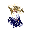
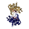
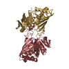
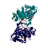
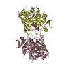
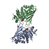
| Title | STUDIES OF ASYMMETRY IN THE THREE-DIMENSIONAL STRUCTURE OF LOBSTER D-GLYCERALDEHYDE-3-PHOSPHATE DEHYDROGENASE | |||||||||
 Components Components | D-GLYCERALDEHYDE-3-PHOSPHATE DEHYDROGENASE | |||||||||
 Keywords Keywords |  OXIDOREDUCTASE / OXIDO-REDUCTASE(ALDEHYDE/DONR / NAD/ACCPT) / OXIDO-REDUCTASE(ALDEHYDE-DONR / NAD-ACCPT) COMPLEX OXIDOREDUCTASE / OXIDO-REDUCTASE(ALDEHYDE/DONR / NAD/ACCPT) / OXIDO-REDUCTASE(ALDEHYDE-DONR / NAD-ACCPT) COMPLEX | |||||||||
| Function / homology |  Function and homology informationglyceraldehyde-3-phosphate dehydrogenase (phosphorylating) / glyceraldehyde-3-phosphate dehydrogenase (NAD+) (phosphorylating) activity / glycolytic process / glucose metabolic process / NAD binding / NADP binding / cytoplasm Function and homology informationglyceraldehyde-3-phosphate dehydrogenase (phosphorylating) / glyceraldehyde-3-phosphate dehydrogenase (NAD+) (phosphorylating) activity / glycolytic process / glucose metabolic process / NAD binding / NADP binding / cytoplasmSimilarity search - Function | |||||||||
| Biological species |  Homarus americanus (American lobster) Homarus americanus (American lobster) | |||||||||
| Method | X-RAY DIFFRACTION / Resolution: 2.9 Å | |||||||||
 Authors Authors | Moras, D. / Olsen, K.W. / Sabesan, M.N. / Buehner, M. / Ford, G.C. / Rossmann, M.G. | |||||||||
 Citation Citation | Journal: J.Biol.Chem. / Year: 1975 Title: Studies of asymmetry in the three-dimensional structure of lobster D-glyceraldehyde-3-phosphate dehydrogenase. Authors: Moras, D. / Olsen, K.W. / Sabesan, M.N. / Buehner, M. / Ford, G.C. / Rossmann, M.G. #1: Journal: J.Mol.Biol. / Year: 1976Title: Anion Binding Sites in the Active Center of D-Glyceraldehyde-3-Phosphate Dehydrogenase Authors: Olsen, K.W. / Garavito, R.M. / Sabesan, M.N. / Rossmann, M.G. #2: Journal: J.Mol.Biol. / Year: 1976Title: Studies on Coenzyme Binding to Glyceraldehyde-3-Phosphate Dehydrogenase Authors: Olsen, K.W. / Garavito, R.M. / Sabesan, M.N. / Rossmann, M.G. #3: Journal: STRUCTURE AND CONFORMATION OF NUCLEIC ACIDS AND PROTEIN-NUCLEIC ACID INTERACTIONS : PROCEEDINGS OF THE FOURTH ANNUAL HARRY STEENBOCK SYMPOSIUM, JUNE 16-19, 1974, MADISON, WISCONSINYear: 1975 Title: A Comparison of the Binding and Function of Nad with Respect to Lactate Dehydrogenase and Glyceraldehyde-3-Phosphate Dehydrogenase Authors: Rossmann, M.G. #4: Journal: J.Biol.Chem. / Year: 1975Title: Sequence Variability and Structure of D-Glyceraldehyde-3-Phosphate Dehydrogenase Authors: Olsen, K.W. / Moras, D. / Rossmann, M.G. / Harris, J.I. #5: Journal: J.Mol.Biol. / Year: 1974Title: Three-Dimensional Structure of D-Glyceraldehyde-3-Phosphate Dehydrogenase Authors: Buehner, M. / Ford, G.C. / Moras, D. / Olsen, K.W. / Rossmann, M.G. #6: Journal: J.Mol.Biol. / Year: 1974Title: Structure Determination of Crystalline Lobster D-Glyceraldehyde-3-Phosphate Dehydrogenase Authors: Buehner, M. / Ford, G.C. / Moras, D. / Olsen, K.W. / Rossmann, M.G. #7: Journal: Proc.Natl.Acad.Sci.USA / Year: 1973Title: D-Glyceraldehyde-3-Phosphate Dehydrogenase,Three Dimensional Structure and Evolutionary Significance Authors: Buehner, M. / Ford, G.C. / Moras, D. / Olsen, K.W. / Rossmann, M.G. #8: Journal: Acta Crystallogr.,Sect.A / Year: 1975Title: An Application of the Molecular Replacement Technique in Direct Space to a Known Protein Structure Authors: Argos, P. / Ford, G.C. / Rossmann, M.G. | |||||||||
| History |
| |||||||||
| Remark 700 | SHEET THE SHEET SUBSTRUCTURE OF THE CATALYTIC DOMAIN IS BIFURCATED. TO REPRESENT THIS FEATURE ...SHEET THE SHEET SUBSTRUCTURE OF THE CATALYTIC DOMAIN IS BIFURCATED. TO REPRESENT THIS FEATURE REDUNDANT SHEETS ARE DEFINED FOR EACH OF THE TWO SUBUNITS. STRANDS 1-7 OF SHEETS GC1 AND RC1 ARE IDENTICAL TO STRANDS 1-7 OF SHEETS GC2 AND RC2. |
- Structure visualization
| Structure viewer | Molecule: MolmilJmol/JSmol |
|---|
- Downloads & links
-Download
| PDBx/mmCIF format | 1gpd.cif.gz | 129.2 KB | Display | PDBx/mmCIF format |
|---|---|---|---|---|
| PDB format | pdb1gpd.ent.gz | 92.2 KB | Display | PDB format |
| PDBx/mmJSON format | 1gpd.json.gz | Tree view | PDBx/mmJSON format | |
| Others |  Other downloads Other downloads |
-Validation report
| Arichive directory | https://data.pdbj.org/pub/pdb/validation_reports/gp/1gpdftp://data.pdbj.org/pub/pdb/validation_reports/gp/1gpd | HTTPS FTP |
|---|
-Related structure data
| Similar structure data |
|---|


-Links
 PDBj
PDBj
- Assembly
| Deposited unit | 
| ||||||||||||||||
|---|---|---|---|---|---|---|---|---|---|---|---|---|---|---|---|---|---|
| 1 | 
| ||||||||||||||||
| Unit cell |
| ||||||||||||||||
| Noncrystallographic symmetry (NCS) | NCS oper:
|
-Components
| #1: Protein | Mass: 35783.949 Da / Num. of mol.: 2 Source method: isolated from a genetically manipulated source Source: (gene. exp.) Homarus americanus (American lobster)References: UniProt: P00357, glyceraldehyde-3-phosphate dehydrogenase (phosphorylating)#2: Chemical | Nicotinamide adenine dinucleotide  Mass: 663.425 Da / Num. of mol.: 2 / Source method: obtained synthetically / Formula: C21H27N7O14P2 / Comment: NAD*YM Mass: 663.425 Da / Num. of mol.: 2 / Source method: obtained synthetically / Formula: C21H27N7O14P2 / Comment: NAD*YM#3: Chemical | ChemComp-PO4 / Phosphate  Mass: 94.971 Da / Num. of mol.: 4 / Source method: obtained synthetically / Formula: PO4 Mass: 94.971 Da / Num. of mol.: 4 / Source method: obtained synthetically / Formula: PO4 |
|---|
-Experimental details
-Experiment
| Experiment | Method: X-RAY DIFFRACTION |
|---|
- Sample preparation
| Crystal | Density Matthews: 5.84 Å3/Da / Density % sol: 78.95 % | ||||||||||||||||||||
|---|---|---|---|---|---|---|---|---|---|---|---|---|---|---|---|---|---|---|---|---|---|
| Crystal grow | *PLUS pH: 5.5 / Method: unknownDetails: (batch), took Watoson & Banaszak from Buehner et al., from original paper. | ||||||||||||||||||||
| Components of the solutions | *PLUS
|
-Data collection
| Radiation | Scattering type: x-ray |
|---|---|
| Radiation wavelength | Relative weight: 1 |
| Reflection | *PLUS Highest resolution: 2.9 Å / Num. obs: 13090 / Num. measured all: 33302 |
- Processing
| Refinement | Highest resolution: 2.9 Å | ||||||||||||
|---|---|---|---|---|---|---|---|---|---|---|---|---|---|
| Refinement step | Cycle: LAST / Highest resolution: 2.9 Å
| ||||||||||||
| Refinement | *PLUS Highest resolution: 2.9 Å | ||||||||||||
| Solvent computation | *PLUS | ||||||||||||
| Displacement parameters | *PLUS |
