 Movie
Movie Controller

[English] 日本語
 Yorodumi
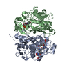
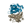
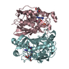
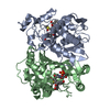
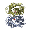
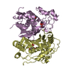
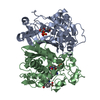
- PDB-1f28: CRYSTAL STRUCTURE OF THYMIDYLATE SYNTHASE FROM PNEUMOCYSTIS CARIN... -
+ Open data

- Basic information
| Entry | Database: PDB / ID: 1f28 | ||||||
|---|---|---|---|---|---|---|---|
| Title | CRYSTAL STRUCTURE OF THYMIDYLATE SYNTHASE FROM PNEUMOCYSTIS CARINII BOUND TO DUMP AND BW1843U89 | ||||||
 Components Components | THYMIDYLATE SYNTHASE | ||||||
 Keywords Keywords | TRANSFERASE / beta-sheet / protein-inhibitor complex | ||||||
| Function / homology |  Function and homology information Function and homology informationphosphopantothenoylcysteine decarboxylase activity / thymidylate synthase / thymidylate synthase activity / dTMP biosynthetic process / dTTP biosynthetic process / protein phosphatase inhibitor activity / nuclear periphery / FMN binding / methylation / mitochondrion / cytosol Similarity search - Function | ||||||
| Biological species |  Pneumocystis carinii (fungus) Pneumocystis carinii (fungus) | ||||||
| Method |  X-RAY DIFFRACTION / SYNCHROTRON / Resolution: 1.9 Å X-RAY DIFFRACTION / SYNCHROTRON / Resolution: 1.9 Å | ||||||
 Authors Authors | Anderson, A.C. / O'Neil, R.H. / Surti, T.S. / Stroud, R.M. | ||||||
 Citation Citation | Journal: Chem.Biol. / Year: 2001 Title: Approaches to solving the rigid receptor problem by identifying a minimal set of flexible residues during ligand docking. Authors: Anderson, A.C. / O'Neil, R.H. / Surti, T.S. / Stroud, R.M. | ||||||
| History |
|
- Structure visualization
| Structure viewer | Molecule: MolmilJmol/JSmol |
|---|
- Downloads & links
-Download
| PDBx/mmCIF format | 1f28.cif.gz | 260.8 KB | Display | PDBx/mmCIF format |
|---|---|---|---|---|
| PDB format | pdb1f28.ent.gz | 210.8 KB | Display | PDB format |
| PDBx/mmJSON format | 1f28.json.gz | Tree view | PDBx/mmJSON format | |
| Others |  Other downloads Other downloads |
-Validation report
| Arichive directory | https://data.pdbj.org/pub/pdb/validation_reports/f2/1f28ftp://data.pdbj.org/pub/pdb/validation_reports/f2/1f28 | HTTPS FTP |
|---|
-Related structure data
| Related structure data | |
|---|---|
| Similar structure data |


-Links
 PDBj
PDBj- Assembly
| Deposited unit | 
| ||||||||
|---|---|---|---|---|---|---|---|---|---|
| 1 | 
| ||||||||
| 2 | 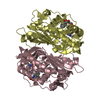
| ||||||||
| Unit cell |
| ||||||||
| Details | The dimer constructed from chains A and B or chains C and D is the biological assembly |
-Components
| #1: Protein | Mass: 34403.281 Da / Num. of mol.: 4 Source method: isolated from a genetically manipulated source Source: (gene. exp.) Pneumocystis carinii (fungus) / Plasmid: PUETS-1.8 / Production host:  #2: Chemical | ChemComp-UMP /   Mass: 308.182 Da / Num. of mol.: 4 / Source method: obtained synthetically / Formula: C9H13N2O8P Mass: 308.182 Da / Num. of mol.: 4 / Source method: obtained synthetically / Formula: C9H13N2O8P#3: Chemical | ChemComp-F89 / 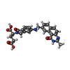  Mass: 500.503 Da / Num. of mol.: 4 / Source method: obtained synthetically / Formula: C27H24N4O6 Mass: 500.503 Da / Num. of mol.: 4 / Source method: obtained synthetically / Formula: C27H24N4O6#4: Water | ChemComp-HOH / |  Mass: 18.015 Da / Num. of mol.: 500 / Source method: isolated from a natural source / Formula: H2O Mass: 18.015 Da / Num. of mol.: 500 / Source method: isolated from a natural source / Formula: H2OHas protein modification | Y | |
|---|
-Experimental details
-Experiment
| Experiment | Method: X-RAY DIFFRACTION / Number of used crystals: 1 |
|---|
- Sample preparation
| Crystal | Density Matthews: 2.41 Å3/Da / Density % sol: 48.95 % | |||||||||||||||||||||||||||||||||||||||||||||
|---|---|---|---|---|---|---|---|---|---|---|---|---|---|---|---|---|---|---|---|---|---|---|---|---|---|---|---|---|---|---|---|---|---|---|---|---|---|---|---|---|---|---|---|---|---|---|
| Crystal grow | Temperature: 297 K / Method: vapor diffusion, hanging drop / pH: 7.4 Details: PEG 8000, Tris, Ammonium sulfate, DTT, pH 7.4, VAPOR DIFFUSION, HANGING DROP, temperature 24K | |||||||||||||||||||||||||||||||||||||||||||||
| Crystal grow | *PLUS | |||||||||||||||||||||||||||||||||||||||||||||
| Components of the solutions | *PLUS
|
-Data collection
| Diffraction | Mean temperature: 137 K |
|---|---|
| Diffraction source | Source: SYNCHROTRON / Site: SSRL  / Beamline: BL9-1 / Wavelength: 1.08 / Beamline: BL9-1 / Wavelength: 1.08 |
| Detector | Type: MARRESEARCH / Detector: IMAGE PLATE / Date: Mar 22, 1999 |
| Radiation | Protocol: SINGLE WAVELENGTH / Monochromatic (M) / Laue (L): M / Scattering type: x-ray |
| Radiation wavelength | Wavelength: 1.08 Å / Relative weight: 1 |
| Reflection | Resolution: 1.9→500 Å / Num. all: 104488 / Num. obs: 94284 / % possible obs: 90.3 % / Observed criterion σ(F): 2 / Observed criterion σ(I): 2 / Redundancy: 4 % / Biso Wilson estimate: 16.3 Å2 / Rmerge(I) obs: 0.088 / Net I/σ(I): 14.4 |
| Reflection shell | Resolution: 1.9→1.93 Å / Redundancy: 4 % / Rmerge(I) obs: 0.523 / Num. unique all: 3882 / % possible all: 75.1 |
| Reflection | *PLUS Lowest resolution: 50 Å / Num. obs: 104488 / Num. measured all: 530911 |
- Processing
| Software |
| ||||||||||||||||||||||||||||||||||||||||
|---|---|---|---|---|---|---|---|---|---|---|---|---|---|---|---|---|---|---|---|---|---|---|---|---|---|---|---|---|---|---|---|---|---|---|---|---|---|---|---|---|---|
| Refinement | Resolution: 1.9→22.4 Å / Rfactor Rfree error: 0.003 / Data cutoff high absF: 2037142.52 / Data cutoff low absF: 0 / Isotropic thermal model: RESTRAINED / Cross valid method: THROUGHOUT / σ(F): 2 / σ(I): 0 / Stereochemistry target values: Engh & Huber
| ||||||||||||||||||||||||||||||||||||||||
| Solvent computation | Solvent model: FLAT MODEL / Bsol: 44.37 Å2 / ksol: 0.357 e/Å3 | ||||||||||||||||||||||||||||||||||||||||
| Displacement parameters | Biso mean: 30.8 Å2
| ||||||||||||||||||||||||||||||||||||||||
| Refine analyze |
| ||||||||||||||||||||||||||||||||||||||||
| Refinement step | Cycle: LAST / Resolution: 1.9→22.4 Å
| ||||||||||||||||||||||||||||||||||||||||
| Refine LS restraints |
| ||||||||||||||||||||||||||||||||||||||||
| Refine LS restraints NCS | NCS model details: CONSTR | ||||||||||||||||||||||||||||||||||||||||
| LS refinement shell | Resolution: 1.9→2.02 Å / Rfactor Rfree error: 0.008 / Total num. of bins used: 6
| ||||||||||||||||||||||||||||||||||||||||
| Xplor file |
| ||||||||||||||||||||||||||||||||||||||||
| Software | *PLUS Name: CNS / Version: 0.9 / Classification: refinement | ||||||||||||||||||||||||||||||||||||||||
| Refinement | *PLUS σ(F): 2 / % reflection Rfree: 10 % | ||||||||||||||||||||||||||||||||||||||||
| Solvent computation | *PLUS | ||||||||||||||||||||||||||||||||||||||||
| Displacement parameters | *PLUS Biso mean: 30.8 Å2 | ||||||||||||||||||||||||||||||||||||||||
| Refine LS restraints | *PLUS
| ||||||||||||||||||||||||||||||||||||||||
| LS refinement shell | *PLUS Rfactor Rfree: 0.279 / % reflection Rfree: 10.3 % / Rfactor Rwork: 0.249 |
