 Movie
Movie Controller

[English] 日本語
 Yorodumi
- PDB-2aaz: Cryptococcus neoformans thymidylate synthase complexed with subst... -
+ Open data

- Basic information
| Entry | Database: PDB / ID: 2aaz | ||||||
|---|---|---|---|---|---|---|---|
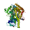
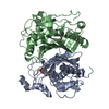
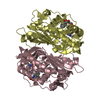
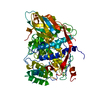
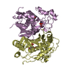
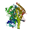
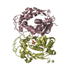
| Title | Cryptococcus neoformans thymidylate synthase complexed with substrate and an antifolate | ||||||
 Components Components | Thymidylate synthase | ||||||
 Keywords Keywords | TRANSFERASE / Methyl transferase / Nucleotide biosynthesis | ||||||
| Function / homology |  Function and homology information Function and homology informationthymidylate synthase / thymidylate synthase activity / dTTP biosynthetic process / dTMP biosynthetic process / methylation / mitochondrion / cytosol Similarity search - Function | ||||||
| Biological species |  Filobasidiella neoformans (Cryptococcus neoformans serotype A) Filobasidiella neoformans (Cryptococcus neoformans serotype A) | ||||||
| Method |  X-RAY DIFFRACTION / SYNCHROTRON / MOLECULAR REPLACEMENT / Resolution: 2.08 Å X-RAY DIFFRACTION / SYNCHROTRON / MOLECULAR REPLACEMENT / Resolution: 2.08 Å | ||||||
 Authors Authors | Finer-Moore, J.S. / Anderson, A.C. / O'Neil, R.H. / Costi, M.P. / Ferrari, S. / Krucinski, J. / Stroud, R.M. | ||||||
 Citation Citation | Journal: Acta Crystallogr.,Sect.D / Year: 2005 Title: The structure of Cryptococcus neoformans thymidylate synthase suggests strategies for using target dynamics for species-specific inhibition. Authors: Finer-Moore, J.S. / Anderson, A.C. / O'Neil, R.H. / Costi, M.P. / Ferrari, S. / Krucinski, J. / Stroud, R.M. #1: Journal: J.Med.Chem. / Year: 1999 Title: Phthalein derivatives as a new tool for selectivity in thymidylate synthase inhibition Authors: Costi, P.M. / Rinaldi, M. / Tondi, D. / Pecorari, P. / Barlocco, D. / Ghelli, S. / Stroud, R.M. / Santi, D.V. / Stout, T.J. / Musiu, C. / Marangiu, E.M. / Pani, A. / Congiu, D. / Loi, G.A. / La Colla, P. #2: Journal: Biochemistry / Year: 1999Title: Structure-based design of inhibitors specific for bacterial thymidylate synthase Authors: Stout, T.J. / Tondi, D. / Rinaldi, M. / Barlocco, D. / Pecorari, P. / Santi, D.V. / Kuntz, I.D. / Stroud, R.M. / Shoichet, B.K. / Costi, M.P. | ||||||
| History |
|
- Structure visualization
| Structure viewer | Molecule: MolmilJmol/JSmol |
|---|
- Downloads & links
-Download
| PDBx/mmCIF format | 2aaz.cif.gz | 3.6 MB | Display | PDBx/mmCIF format |
|---|---|---|---|---|
| PDB format | pdb2aaz.ent.gz | 3.1 MB | Display | PDB format |
| PDBx/mmJSON format | 2aaz.json.gz | Tree view | PDBx/mmJSON format | |
| Others |  Other downloads Other downloads |
-Validation report
| Arichive directory | https://data.pdbj.org/pub/pdb/validation_reports/aa/2aazftp://data.pdbj.org/pub/pdb/validation_reports/aa/2aaz | HTTPS FTP |
|---|
-Related structure data


-Links
 PDBj
PDBj- Assembly
| Deposited unit | 
| ||||||||
|---|---|---|---|---|---|---|---|---|---|
| 1 | 
| ||||||||
| 2 | 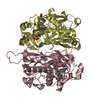
| ||||||||
| 3 | 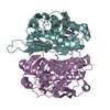
| ||||||||
| 4 | 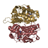
| ||||||||
| 5 | 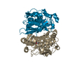
| ||||||||
| 6 | 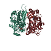
| ||||||||
| 7 | 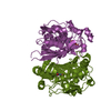
| ||||||||
| 8 | 
| ||||||||
| Unit cell |
| ||||||||
| Number of models | 4 | ||||||||
| Components on special symmetry positions |
| ||||||||
| Details | The biological assembly is a dimer. There are 8 dimers in the asymmetric unit and four of these dimers are 4-fold statistically disordered. Pairs of chains that constitute the four fully-occupied biological dimers are (A,B), (C,D), (E,F) and (G,H). Dimers that are in four 1/4-occupied packing arrangements in the cell are chains (I,J), (K,L), (M,N) and (O,P) |

-Components
| #1: Protein | Mass: 35911.977 Da / Num. of mol.: 16 Source method: isolated from a genetically manipulated source Source: (gene. exp.) Filobasidiella neoformans (Cryptococcus neoformans serotype A)Gene: TMP1 / Plasmid: pET CNTS / Species (production host): Escherichia coli / Production host:  References: UniProt: P45351, UniProt: P0CS12*PLUS, thymidylate synthase #2: Chemical | ChemComp-UMP /   Mass: 308.182 Da / Num. of mol.: 16 / Source method: obtained synthetically / Formula: C9H13N2O8P Mass: 308.182 Da / Num. of mol.: 16 / Source method: obtained synthetically / Formula: C9H13N2O8P#3: Chemical | ChemComp-CB3 /   Mass: 477.469 Da / Num. of mol.: 8 / Source method: obtained synthetically / Formula: C24H23N5O6 Mass: 477.469 Da / Num. of mol.: 8 / Source method: obtained synthetically / Formula: C24H23N5O6#4: Water | ChemComp-HOH / |  Mass: 18.015 Da / Num. of mol.: 1212 / Source method: isolated from a natural source / Formula: H2O Mass: 18.015 Da / Num. of mol.: 1212 / Source method: isolated from a natural source / Formula: H2OHas protein modification | Y | |
|---|
-Experimental details
-Experiment
| Experiment | Method: X-RAY DIFFRACTION / Number of used crystals: 1 |
|---|
- Sample preparation
| Crystal | Density Matthews: 2.93 Å3/Da / Density % sol: 57.99 % |
|---|---|
| Crystal grow | Temperature: 293 K / Method: vapor diffusion, hanging drop / pH: 8.4 Details: 0.107 mM protein, dUMP,CB3, DTT, PEG4000/PEG6000, Tris, Na acetate/K acetate, pH 8.4, VAPOR DIFFUSION, HANGING DROP, temperature 293K |
-Data collection
| Diffraction | Mean temperature: 103 K |
|---|---|
| Diffraction source | Source: SYNCHROTRON / Site: SSRL  / Beamline: BL9-1 / Wavelength: 1 Å / Beamline: BL9-1 / Wavelength: 1 Å |
| Detector | Type: MARRESEARCH / Detector: IMAGE PLATE / Date: May 19, 2000 / Details: monochrometer |
| Radiation | Protocol: SINGLE WAVELENGTH / Monochromatic (M) / Laue (L): M / Scattering type: x-ray |
| Radiation wavelength | Wavelength: 1 Å / Relative weight: 1 |
| Reflection | Resolution: 2.07→99 Å / Num. all: 389001 / Num. obs: 389001 / % possible obs: 97 % / Observed criterion σ(F): -3 / Observed criterion σ(I): -3 / Redundancy: 1.85 % / Biso Wilson estimate: 23.2 Å2 / Rmerge(I) obs: 0.1 / Net I/σ(I): 7.4 |
| Reflection shell | Resolution: 2.07→2.14 Å / Rmerge(I) obs: 0.344 / Mean I/σ(I) obs: 1.7 / % possible all: 97.9 |
- Processing
| Software |
| ||||||||||||||||||||||||||||||||||||
|---|---|---|---|---|---|---|---|---|---|---|---|---|---|---|---|---|---|---|---|---|---|---|---|---|---|---|---|---|---|---|---|---|---|---|---|---|---|
| Refinement | Method to determine structure: MOLECULAR REPLACEMENT / Resolution: 2.08→49.91 Å / Rfactor Rfree error: 0.002 / Data cutoff high absF: 5580798.34 / Data cutoff low absF: 0 / Isotropic thermal model: RESTRAINED / Cross valid method: THROUGHOUT / σ(F): 0 / Stereochemistry target values: Engh & Huber Details: Tight NCS restraints used throughout refinement. Structures of all monomers in the asymmetric unit restrained to be equivalent except for the following C. neoformans thymidylate synthase ...Details: Tight NCS restraints used throughout refinement. Structures of all monomers in the asymmetric unit restrained to be equivalent except for the following C. neoformans thymidylate synthase inserts: residues 13-16, 107-108 200-202. The following residues not present in the model: 1-12 for chains A-H and side chain of K107 in chains A and C, residues 1-15 and 200-202 in chains I-P.
| ||||||||||||||||||||||||||||||||||||
| Solvent computation | Solvent model: FLAT MODEL / Bsol: 49.4499 Å2 / ksol: 0.20613 e/Å3 | ||||||||||||||||||||||||||||||||||||
| Displacement parameters | Biso mean: 28.5 Å2
| ||||||||||||||||||||||||||||||||||||
| Refine analyze |
| ||||||||||||||||||||||||||||||||||||
| Refinement step | Cycle: LAST / Resolution: 2.08→49.91 Å
| ||||||||||||||||||||||||||||||||||||
| Refine LS restraints |
| ||||||||||||||||||||||||||||||||||||
| Refine LS restraints NCS | NCS model details: CONSTR | ||||||||||||||||||||||||||||||||||||
| LS refinement shell | Resolution: 2.08→2.21 Å / Rfactor Rfree error: 0.005 / Total num. of bins used: 6
| ||||||||||||||||||||||||||||||||||||
| Xplor file |
|
