 Movie
Movie Controller

[English] 日本語
 Yorodumi
- PDB-1ebl: THE 1.8 A CRYSTAL STRUCTURE AND ACTIVE SITE ARCHITECTURE OF BETA-... -
+ Open data

- Basic information
| Entry | Database: PDB / ID: 1ebl | ||||||
|---|---|---|---|---|---|---|---|
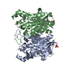
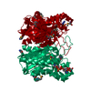
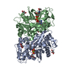
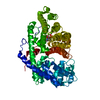
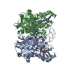
| Title | THE 1.8 A CRYSTAL STRUCTURE AND ACTIVE SITE ARCHITECTURE OF BETA-KETOACYL-[ACYL CARRIER PROTEIN] SYNTHASE III (FABH) FROM ESCHERICHIA COLI | ||||||
 Components Components | BETA-KETOACYL-ACP SYNTHASE III | ||||||
 Keywords Keywords | TRANSFERASE / ACYLTRANSFERASE / CONDENSING ENZYME / FATTY ACID SYNTHESIS / LIPID METABOLISM / ALPHA-BETA PROTEIN / FIVE-LAYERED FOLD / COENZYME A BINDING PROTEIN / HELIX DIPOLE / MALONYL COA DECARBOXYLATING ENZYME | ||||||
| Function / homology |  Function and homology information Function and homology informationbeta-ketoacyl-[acyl-carrier-protein] synthase III / beta-ketoacyl-acyl-carrier-protein synthase III activity / 3-oxoacyl-[acyl-carrier-protein] synthase activity / fatty acid metabolic process / fatty acid biosynthetic process / cytosolSimilarity search - Function | ||||||
| Biological species |  Escherichia coli (E. coli) Escherichia coli (E. coli) | ||||||
| Method | X-RAY DIFFRACTION / SYNCHROTRON / Resolution: 1.8 Å | ||||||
 Authors Authors | Davies, C. / Heath, R.J. / White, S.W. / Rock, C.O. | ||||||
 Citation Citation | Journal: Structure Fold.Des. / Year: 2000 Title: The 1.8 A crystal structure and active-site architecture of beta-ketoacyl-acyl carrier protein synthase III (FabH) from escherichia coli. Authors: Davies, C. / Heath, R.J. / White, S.W. / Rock, C.O. #1: Journal: J.Biol.Chem. / Year: 1992Title: Isolation and characterization of the beta-ketoacyl-acyl carrier protein synthase III gene (fabH) from Escherichia coli K-12 Authors: Tsay, J.T. / Oh, W. / Larson, T.J. / Jackowski, S. / Rock, C.O. | ||||||
| History |
|
- Structure visualization
| Structure viewer | Molecule: MolmilJmol/JSmol |
|---|
- Downloads & links
-Download
| PDBx/mmCIF format | 1ebl.cif.gz | 144.8 KB | Display | PDBx/mmCIF format |
|---|---|---|---|---|
| PDB format | pdb1ebl.ent.gz | 119.4 KB | Display | PDB format |
| PDBx/mmJSON format | 1ebl.json.gz | Tree view | PDBx/mmJSON format | |
| Others |  Other downloads Other downloads |
-Validation report
| Arichive directory | https://data.pdbj.org/pub/pdb/validation_reports/eb/1eblftp://data.pdbj.org/pub/pdb/validation_reports/eb/1ebl | HTTPS FTP |
|---|
-Related structure data
| Related structure data | |
|---|---|
| Similar structure data |


-Links
 PDBj
PDBj


- Assembly
| Deposited unit | 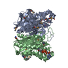
| ||||||||
|---|---|---|---|---|---|---|---|---|---|
| 1 |
| ||||||||
| Unit cell |
| ||||||||
| Noncrystallographic symmetry (NCS) | NCS oper: (Code: given Matrix: (-0.33103, -0.40231, -0.85256), Vector : Details | The biological assembly is a dimer constructed from chain A and chain B as represented in the crystal asymmetric unit. | |
-Components
| #1: Protein | / 3-OXOACYL-[ACYL-CARRIER-PROTEIN] SYNTHASE III / KAS III Mass: 33923.129 Da / Num. of mol.: 2 Source method: isolated from a genetically manipulated source Source: (gene. exp.) Escherichia coli (E. coli) / Plasmid: PET-15B / Production host: Escherichia coli (E. coli)References: UniProt: P0A6R0, beta-ketoacyl-[acyl-carrier-protein] synthase I #2: Chemical | Coenzyme A  Mass: 767.534 Da / Num. of mol.: 2 / Source method: obtained synthetically / Formula: C21H36N7O16P3S Mass: 767.534 Da / Num. of mol.: 2 / Source method: obtained synthetically / Formula: C21H36N7O16P3S#3: Water | ChemComp-HOH / | Water Mass: 18.015 Da / Num. of mol.: 704 / Source method: isolated from a natural source / Formula: H2O Mass: 18.015 Da / Num. of mol.: 704 / Source method: isolated from a natural source / Formula: H2O |
|---|
-Experimental details
-Experiment
| Experiment | Method: X-RAY DIFFRACTION / Number of used crystals: 1 |
|---|
- Sample preparation
| Crystal | Density Matthews: 2.48 Å3/Da / Density % sol: 50.42 % | ||||||||||||||||||||||||||||||||||||||||
|---|---|---|---|---|---|---|---|---|---|---|---|---|---|---|---|---|---|---|---|---|---|---|---|---|---|---|---|---|---|---|---|---|---|---|---|---|---|---|---|---|---|
| Crystal grow | Temperature: 298 K / Method: vapor diffusion, hanging drop / pH: 7.5 Details: 1.8-2.0 M ammonium sulphate, 2% PEG 400, 0.1M HEPES, pH 7.5, VAPOR DIFFUSION, HANGING DROP, temperature 298.0K | ||||||||||||||||||||||||||||||||||||||||
| Crystal | *PLUS Density % sol: 50.5 % | ||||||||||||||||||||||||||||||||||||||||
| Crystal grow | *PLUS pH: 7.4 | ||||||||||||||||||||||||||||||||||||||||
| Components of the solutions | *PLUS
|
-Data collection
| Diffraction | Mean temperature: 100 K |
|---|---|
| Diffraction source | Source: SYNCHROTRON / Site: CHESS  / Beamline: F2 / Wavelength: 1 / Beamline: F2 / Wavelength: 1 |
| Detector | Type: OTHER / Detector: CCD / Date: Jan 21, 1999 |
| Radiation | Protocol: SINGLE WAVELENGTH / Monochromatic (M) / Laue (L): M / Scattering type: x-ray |
| Radiation wavelength | Wavelength: 1 Å / Relative weight: 1 |
| Reflection | Resolution: 1.8→31.6 Å / Num. all: 230489 / Num. obs: 61752 / % possible obs: 97.4 % / Observed criterion σ(F): 0 / Observed criterion σ(I): 0 / Redundancy: 3.73 % / Biso Wilson estimate: 17.17 Å2 / Rmerge(I) obs: 0.056 / Net I/σ(I): 20.52 |
| Reflection shell | Resolution: 1.8→1.89 Å / Redundancy: 2.88 % / Rmerge(I) obs: 0.14 / Num. unique all: 8193 / % possible all: 89.8 |
| Reflection | *PLUS Num. measured all: 230489 |
| Reflection shell | *PLUS % possible obs: 89.8 % |
- Processing
| Software |
| |||||||||||||||||||||||||
|---|---|---|---|---|---|---|---|---|---|---|---|---|---|---|---|---|---|---|---|---|---|---|---|---|---|---|
| Refinement | Resolution: 1.8→20 Å / σ(F): 0 / σ(I): 0 / Stereochemistry target values: Engh & Huber Details: Restrained refinement with maximum likelihood residual, Minimization by sparse matrix
| |||||||||||||||||||||||||
| Refinement step | Cycle: LAST / Resolution: 1.8→20 Å
| |||||||||||||||||||||||||
| Refine LS restraints |
| |||||||||||||||||||||||||
| Software | *PLUS Name: REFMAC / Classification: refinement | |||||||||||||||||||||||||
| Refine LS restraints | *PLUS
|
